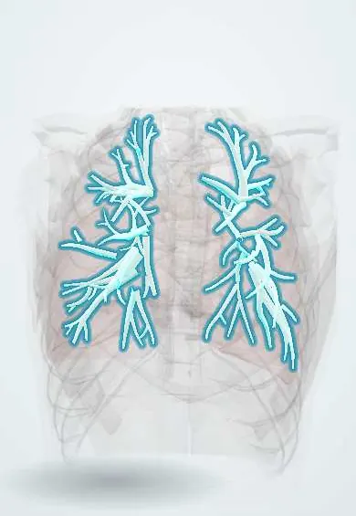
Bronchiectasis !
Bronchiectasis is a permanent widening of the airways that traps mucus, causing a long-lasting cough with phlegm and repeated chest infections.
Updated 2026-10-30
Structures involved
Tap one to see what it is and open it in 3D.
Microorganisms that cause it
In histology
- Electron microscopy: what it shows that light can't
- Cilia, microvilli and stereocilia
- The airway: from nose to bronchioles
In the clinical exam
- Digital clubbing and nail signs
- Adventitious sounds: crackles, wheezes, rhonchi, stridor and pleural rub
- Respiratory symptoms: cough, sputum, hemoptysis and dyspnea
In physiology
- Obstructive and restrictive patterns, and flow–volume loops
- Airway resistance
- Airway defenses, cough and mucociliary clearance
- The bronchial circulation
Why it happens
- Cystic fibrosis: from the CFTR channel to organ damage
- When leukocytes injure tissue: proteases, oxidants and NETs
What it is
Permanent, abnormal widening of the bronchi with chronic inflammation and poor mucus clearance, causing a long-term productive cough and repeated chest infections.
Who gets it
Increasingly recognised with CT, more often in women and older adults. In Latin America past tuberculosis and childhood infections are key causes; cystic fibrosis is the classic cause in young people. In six cohorts from Argentina, Brazil and Chile (651 patients, mean age 48, about two-thirds women), 14.6% died within five years.
Causes
- Post-infectious: tuberculosis, severe pneumonia, whooping cough, measles
- Cystic fibrosis
- Primary ciliary dyskinesia
- Immunodeficiency
- Allergic bronchopulmonary aspergillosis
- Connective tissue disease, COPD, airway obstruction; many cases are idiopathic
Risk factors
- Previous tuberculosis or severe childhood lung infection
- Cystic fibrosis or other genetic disease
- Immune deficiency
- Rheumatoid arthritis or inflammatory bowel disease
- Aspiration
- Severe asthma or COPD
Pathophysiology
An initial insult damages the airway wall, and a self-perpetuating cycle follows: impaired mucus clearance, chronic bacterial infection and neutrophil inflammation that destroys elastic and muscle tissue, so the bronchi dilate further. Pseudomonas colonisation marks more severe disease. Enlarged bronchial arteries can bleed.
Classification
- Cylindrical
- Varicose
- Cystic (saccular)
- Focal or diffuse
- Cystic fibrosis or non-cystic fibrosis bronchiectasis
Symptoms and signs
- Chronic cough with daily purulent sputum
- Repeated chest infections (exacerbations)
- Breathlessness and wheeze
- Haemoptysis
- Fatigue
- Coarse crackles; clubbing in advanced disease
Diagnosis
- Clinical
- Chronic productive cough and recurrent infections; the cause is then sought.
- Tests
- Sputum culture including mycobacteria; spirometry (often obstructive); blood count, immunoglobulins and tests for aspergillosis; sweat test or genetic tests when cystic fibrosis is suspected.
- Imaging
- High-resolution CT confirms it: bronchus wider than its artery (signet ring sign), lack of tapering and visible airways near the pleura. Chest X-ray may show tram-track lines.
Differential diagnosis
- COPD
- Asthma
- Pulmonary tuberculosis
- Chronic bronchitis
- Lung cancer
Goal of treatment
Clear mucus, reduce exacerbations, treat the underlying cause and preserve lung function and quality of life.
Treatment
- Rehabilitation
- Daily airway clearance with physiotherapy techniques and devices, and pulmonary rehabilitation.
- Medical
- Antibiotics for exacerbations guided by sputum culture; long-term macrolides or inhaled antibiotics for frequent exacerbations or Pseudomonas.
- Medical
- Treatment of the cause: immunoglobulin replacement, treatment of aspergillosis or mycobacteria, specific cystic fibrosis therapy; mucoactive agents such as hypertonic saline.
- Lifestyle
- Influenza and pneumococcal vaccination, smoking cessation and good nutrition.
- Surgical
- Surgical resection for localised disease that does not respond to medical treatment.
- Procedure
- Bronchial artery embolisation for serious haemoptysis.
Complications
- Recurrent exacerbations and pneumonia
- Massive haemoptysis
- Respiratory failure
- Pulmonary hypertension and cor pulmonale
- Chronic Pseudomonas infection
Prognosis
A chronic condition with variable course. Frequent exacerbations, Pseudomonas, extensive disease and low lung function predict a worse outcome; good airway clearance improves quality of life.
Prevention
- Childhood vaccination (measles, whooping cough)
- Early diagnosis and full treatment of tuberculosis
- Influenza and pneumococcal vaccination
Test yourself
Think of the answer, then tap the question to see it.
What is the imaging test of choice to diagnose bronchiectasis?
High-resolution chest CT, showing a bronchus wider than its artery and airways that do not taper.
What is the signet ring sign?
On CT, a dilated bronchus next to its smaller pulmonary artery, like a ring with a stone.
What are two important causes of bronchiectasis in Latin America?
Previous tuberculosis and severe childhood lung infections; cystic fibrosis is the classic cause in young people.
Which organism colonising the airways signals worse prognosis in bronchiectasis?
Pseudomonas aeruginosa.
How is serious haemoptysis from bronchiectasis usually controlled?
Bronchial artery embolisation, since the bleeding comes from enlarged bronchial arteries.
Related conditions
- Pulmonary tuberculosis
- Chronic obstructive pulmonary disease (COPD)
- Asthma
- Community-acquired pneumonia
- Rheumatoid arthritis
- Lung cancer
Educational information, not medical advice. For any symptom, see a health professional.
Sources: Bronchiectasis (StatPearls (NCBI Bookshelf), 2023); Latin America validation of FACED score in patients with bronchiectasis: an analysis of six cohorts (BMC pulmonary medicine, 2017) · adapted, CC BY 4.0.
Spotted a mistake?We are always improving for the community. Tell us what is wrong and our team will review it.Report a mistake
Representative model, for educational purposes only.