Conditions · Pediatrics and neonatology
Biliary atresia !
In some newborns the bile ducts scar and close, so bile cannot leave the liver. Jaundice beyond two weeks with pale stools needs urgent checks.
Updated 2026-11-01
Structures involved
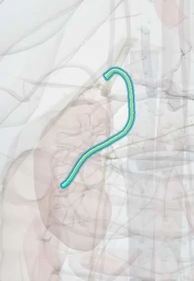
Tap one to see what it is and open it in 3D.
Why it happens
- Fibrosis in organs: TGF-β and the myofibroblast
- Bile and the enterohepatic circulation
- Heme breakdown and bilirubin
- Gallbladder and bile ducts
What it is
A progressive inflammatory and fibrosing obliteration of the extrahepatic bile ducts in infancy, causing obstructive cholestasis and, without treatment, biliary cirrhosis. It is the most common cause of neonatal cholestatic jaundice needing surgery and the leading reason for liver transplantation in children.
Who gets it
Occurs in about 1 in 10,000–20,000 live births, more often in East Asia, and slightly more in girls. Outcomes depend on surgery within the first two months, so stool colour cards and early checks of prolonged jaundice are promoted in Latin America, including Colombia, where late referral remains a problem.
Causes
- Cause unknown; likely an immune-mediated injury to bile ducts after a perinatal trigger
- Proposed triggers: viral infection (e.g. cytomegalovirus, reovirus, rotavirus), toxins, genetic susceptibility
- Syndromic form with other anomalies: polysplenia, situs inversus, preduodenal portal vein, interrupted inferior vena cava, heart defects
Risk factors
- East Asian ancestry
- Female sex (slight)
- Laterality and splenic malformations (syndromic form)
- Maternal diabetes (reported association)
Pathophysiology
Inflammation destroys the extrahepatic ducts, which become fibrous cords, so conjugated bilirubin and bile acids back up into the liver. Without bile in the gut, stools turn pale and fat and fat-soluble vitamins (A, D, E, K) are poorly absorbed, which can cause bleeding from vitamin K deficiency. Ongoing cholestasis drives portal fibrosis that progresses to cirrhosis, portal hypertension and liver failure within the first years of life.
Classification
- Type I: atresia of the common bile duct
- Type II: atresia up to the common hepatic duct
- Type III: atresia of the right and left hepatic ducts at the porta hepatis (most common)
- Isolated, syndromic (biliary atresia splenic malformation) or cystic variants
Symptoms and signs
- Jaundice lasting beyond 2 weeks of age (3 weeks in preterm infants), often after a brief normal interval
- Pale, clay-coloured or white stools (acholia)
- Dark urine that stains the nappy
- Firm, enlarged liver; later splenomegaly
- Initially good feeding and growth, then poor weight gain and irritability
- Abdominal distension from ascites in advanced disease
Diagnosis
- Clinical
- Check fractionated bilirubin in every infant jaundiced beyond 2 weeks; look at the stool colour (stool colour card) and urine. Early referral to a paediatric hepatology centre is essential.
- Tests
- Raised conjugated bilirubin and gamma-glutamyl transferase, raised transaminases; coagulation profile; screening for other causes (alpha-1 antitrypsin, thyroid, infections, metabolic disorders). Liver biopsy shows bile duct proliferation, bile plugs and portal fibrosis. Intraoperative cholangiography confirms the diagnosis.
- Imaging
- Abdominal ultrasound after fasting: absent or small gallbladder, triangular cord sign at the porta hepatis, and polysplenia in the syndromic form. Hepatobiliary scintigraphy shows good uptake but no excretion into the bowel.
Differential diagnosis
- Physiological or breast-milk jaundice (unconjugated)
- Neonatal hepatitis (infectious or idiopathic)
- Alagille syndrome and progressive familial intrahepatic cholestasis
- Alpha-1 antitrypsin deficiency
- Choledochal cyst
- Congenital hypothyroidism, galactosaemia and other metabolic causes
Goal of treatment
Restore bile flow as early as possible to slow liver damage, support nutrition and vitamin absorption, treat complications, and plan liver transplantation when the liver fails.
Treatment
- Surgical
- Kasai portoenterostomy: removal of the fibrotic ducts and joining a Roux-en-Y loop of jejunum to the porta hepatis; best results when done before about 60 days of age.
- Medical
- Nutrition with medium-chain-triglyceride formula and supplements of fat-soluble vitamins A, D, E and K.
- Medical
- Medicines after surgery as directed by the hepatology team: antibiotic prophylaxis against cholangitis, choleretic bile acids, and in some centres corticosteroids.
- Surgical
- Liver transplantation for failed Kasai, late diagnosis with cirrhosis, or progressive liver failure.
- Follow-up
- Nursing monitoring: stool colour, jaundice, temperature (cholangitis), weight and growth, bleeding signs, and vaccination before transplant.
Complications
- Ascending cholangitis after Kasai
- Biliary cirrhosis and liver failure
- Portal hypertension with varices, splenomegaly and ascites
- Malnutrition and fat-soluble vitamin deficiencies (rickets, bleeding)
- Hepatopulmonary syndrome and hepatocellular carcinoma (late)
Prognosis
Untreated, most children die of liver failure within the first years. An early Kasai restores bile flow in about half of infants and a sizeable minority keep their native liver into adulthood; most eventually need a transplant, with good long-term survival after it.
Prevention
- No primary prevention is known
- Screening with stool colour cards and checking conjugated bilirubin in prolonged jaundice allows early surgery
Test yourself
Think of the answer, then tap the question to see it.
What is the classic triad of biliary atresia in an infant?
Prolonged jaundice, pale (acholic) stools and dark urine.
What type of hyperbilirubinaemia is found in biliary atresia?
Conjugated (direct) hyperbilirubinaemia.
What operation restores bile flow in biliary atresia?
The Kasai portoenterostomy.
Why is surgery best done before about 60 days of age?
Earlier surgery gives better bile drainage before fibrosis progresses to cirrhosis.
What is the most common indication for liver transplantation in children?
Biliary atresia.
Related conditions
- Neonatal jaundice
- Cirrhosis
- Portal hypertension
- Vitamin K deficiency bleeding of the newborn
- Hepatocellular carcinoma
Educational information, not medical advice. For any symptom, see a health professional.
Sources: Biliary atresia: MedlinePlus Medical Encyclopedia (MedlinePlus (U.S. National Library of Medicine), 2024).
Spotted a mistake?We are always improving for the community. Tell us what is wrong and our team will review it.Report a mistake
Representative model, for educational purposes only.