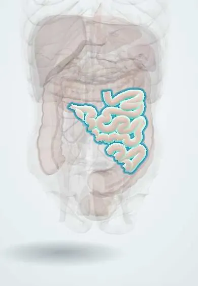
Jejunum !
The jejunum is the middle part of the small intestine, mostly in the upper left belly, where much of the food is absorbed.
Opens in the Durgical app, no sign-up needed.
What it is
The middle part of the small intestine, in the upper left abdomen, with a thick, folded lining.
What it does
Absorbs most nutrients.
Key facts
- Where
- Proximal two-fifths of small intestine from duodenojejunal flexure (left of L2); mostly left upper abdomen; intraperitoneal on mesentery
- Blood supply
- Jejunal branches of SMA: few (1–2) arcades and long vasa recta
- Nerve
- Vagus (parasympathetic); sympathetic via superior mesenteric plexus (T8–T10)
- Function
- Main site of nutrient absorption; thick red wall with tall, dense circular folds
Relations
- Course
- From the duodenojejunal flexure left of L2 its coils lie mainly in the upper left and central abdomen, hung from the mesentery, whose root runs obliquely from left of L2 down to the right sacroiliac joint
- In front
- Greater omentum and the anterior abdominal wall
- Above
- Transverse colon and transverse mesocolon: the jejunum lies in the infracolic compartment, mostly left of the root of the mesentery
- Lateral
- Descending colon; at the duodenojejunal flexure the inferior mesenteric vein runs in a peritoneal fold to its left, beside the paraduodenal fossae
How to find it
Not palpable. At laparotomy the transverse colon is lifted and the first jejunal loop is found emerging from the duodenojejunal flexure, just left of the root of the mesentery under the transverse mesocolon; the bowel is then run distally to the cecum. Held to the light, its mesentery shows few arcades and long vasa recta.
On imaging
On X-ray and CT, jejunal loops in the upper left abdomen show a feathery pattern from dense circular folds that cross the whole width of the lumen, unlike colonic haustral folds, which only partly cross it. Dilated loops with air-fluid levels suggest small-bowel obstruction.
Common problems
- Obstruction from adhesions or hernias.
- Crohn disease (often in the last part of the ileum).
- Celiac disease (gluten damages the lining).
How it is diagnosed
Examination, X-ray or CT, endoscopy, blood tests.
Treatment
- Depends on the cause: rest and fluids for many obstructions, medicines for Crohn disease, surgery for some.
Clinical notes
- Pain from the jejunum (midgut) is felt around the umbilicus
- The proximal jejunum, with its long mobile mesentery, is used for feeding jejunostomies and Roux-en-Y loops in biliary, gastric and bariatric surgery
- Paraduodenal fossae near the duodenojejunal flexure are a site of internal hernia
Development
Midgut: the gut loop herniates into the umbilical cord around week 6, rotates 270° counterclockwise around the superior mesenteric artery and returns by about week 10; malrotation can lead to midgut volvulus in the newborn.
Related conditions
- Small bowel obstruction
- Coeliac disease
- Acute (viral) gastroenteritis
- Acute mesenteric ischaemia
- Hartnup disease
- Lactose intolerance
- Abetalipoproteinemia (Bassen–Kornzweig syndrome)
- Megaloblastic anaemia (B12 or folate deficiency)
- Paralytic ileus and colonic pseudo-obstruction (Ogilvie)
- Dumping syndrome (rapid gastric emptying)
- Small intestinal bacterial overgrowth (SIBO)
- Whipple disease
- Short bowel syndrome
- Tropical sprue
- Gastrointestinal stromal tumour (GIST)
- Soil-transmitted helminthiasis (ascariasis, hookworm, trichuriasis)
- Giardiasis
- Strongyloidiasis
- Cholera
- Cryptosporidiosis and other intestinal coccidia (Cyclospora, Cystoisospora)
- Intestinal tapeworm infections (taeniasis, hymenolepiasis, diphyllobothriasis)
- Dehydration in children (acute diarrhoeal disease)
- Gastroschisis and omphalocele
- Duodenal and intestinal atresia
- Failure to thrive (faltering growth)
- Selective IgA deficiency
- Postoperative ileus
Test yourself
Think of the answer, then tap the question to see it.
How is the jejunum told from the ileum at surgery?
Jejunum: thicker, redder wall, wider lumen, tall dense circular folds, few arcades with long vasa recta, little mesenteric fat, few Peyer patches.
Which landmark marks the start of the jejunum and divides upper from lower GI bleeding?
The duodenojejunal flexure, left of L2, held by the suspensory muscle of the duodenum (ligament of Treitz).
Where is pain from the jejunum felt, and why?
Around the umbilicus, because the jejunum is a midgut organ.
Educational information, not medical advice. For any symptom, see a health professional.
Its names
- Official Latin (TA2)
- jejunum TA2 2958
- Official Spanish (DeCS)
- Yeyuno DeCS D007583
- English (TA2)
- Jejunum
- Also called
- intestinum jejunum
Related
Sources: Terminologia Anatomica (TA2); DeCS; Anatomy, Abdomen and Pelvis, Small Intestine.
Spotted a mistake?We are always improving for the community. Tell us what is wrong and our team will review it.Report a mistake
Representative model, for educational purposes only.