
Brown syndrome
The superior oblique tendon cannot slide freely, so the eye cannot look up when turned towards the nose. Often congenital and mild.
What it is
Brown syndrome is a restrictive strabismus caused by limited movement of the superior oblique tendon through the trochlea. The affected eye cannot elevate in adduction, with better elevation in abduction. It can be congenital or acquired from inflammation, trauma or surgery, and the forced duction test is positive.
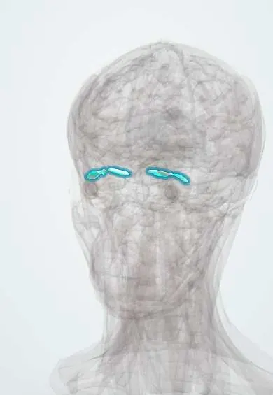
Who gets it
Uncommon; usually congenital, unilateral and more often on the right; bilateral in about one in ten.
Causes
- Congenital: short or inelastic superior oblique tendon-trochlea complex
- Acquired inflammatory: rheumatoid arthritis, juvenile idiopathic arthritis, lupus, sinusitis
- Trauma to the superior nasal orbit
- After superior oblique tuck, sinus or orbital surgery
Risk factors
- Inflammatory arthritis
- Trauma or surgery near the trochlea
Pathophysiology
The superior oblique tendon cannot lengthen or slide through the trochlea when the inferior oblique contracts, mechanically tethering the eye in adduction. Downgaze is normal and superior oblique overaction is absent, which separates it from inferior oblique palsy.
Classification
- Congenital or acquired
- Mild, moderate or severe (by deviation in primary gaze and head posture)
Symptoms and signs
- Eye fails to look up when turned towards the nose
- Chin-up head posture or face turn
- Click or pain near the inner upper orbit (acquired forms)
- Double vision in upgaze
Diagnosis
- Clinical
- Limited elevation in adduction, improved in abduction, widening of the palpebral fissure on adduction, V-pattern divergence in upgaze and no superior oblique overaction.
- Tests
- Positive forced duction test (restriction on elevating the adducted eye); inflammatory and rheumatology tests in acquired cases.
- Imaging
- MRI or CT of the orbit for acquired cases to look at the trochlea and superior oblique tendon.
Differential diagnosis
- Inferior oblique palsy (positive head tilt, superior oblique overaction, A pattern)
- Orbital floor fracture
- Thyroid eye disease
- Monocular elevation deficiency
Eye that will not rise: Brown syndrome vs its look-alikes
| Feature | Brown syndrome | Inferior oblique palsy | Orbital floor fracture | Monocular elevation deficiency |
|---|---|---|---|---|
| Where elevation fails | Mainly in adduction | Mainly in adduction | In upgaze, often also downgaze | Equally in adduction and abduction |
| Forced duction | Restricted on elevating the adducted eye | Free | Restricted on elevation | Restricted or free, depending on type |
| Superior oblique overaction | Absent | Present | Absent | Absent |
| Pattern in up- and downgaze | V pattern (divergence in upgaze) | A pattern | None typical | None typical |
| Other clues | Congenital, or arthritis, trauma or surgery near the trochlea; click | Positive head tilt test | Trauma, cheek numbness, sunken eye | Congenital; may cause pseudoptosis |
Goal of treatment
Allow binocular vision without abnormal head posture; many cases resolve without surgery.
Treatment
- Follow-up
- Observation; congenital and acquired cases may improve spontaneously.
- Medical
- Oral anti-inflammatories or a local corticosteroid injection near the trochlea for inflammatory cases.
- Surgical
- Superior oblique tendon lengthening (spacer, split tendon or tenotomy) for hypotropia in primary gaze or a marked head posture.
- Conservative
- Amblyopia treatment when needed.
Complications
- Amblyopia
- Superior oblique palsy after tendon surgery
- Persistent head posture
Common traps
- Limited elevation in adduction means inferior oblique palsy: Brown syndrome is mechanical, so forced duction is restricted and there is no superior oblique overaction; inferior oblique palsy has free ductions, superior oblique overaction and an A pattern
- A V pattern proves inferior oblique overaction: in Brown syndrome the eyes diverge in upgaze because the tethered eye is pulled outward as it tries to rise, not because the inferior oblique overacts
- Every congenital case needs surgery: many improve with observation; surgery is kept for hypotropia in primary gaze or a marked head posture, because lengthening the tendon can leave a superior oblique palsy
- Acquired Brown syndrome is always traumatic: inflammation at the trochlea from rheumatoid or juvenile idiopathic arthritis, lupus or sinusitis is a classic cause and may respond to anti-inflammatory treatment
- A click near the inner upper orbit is unrelated: a felt or palpable click on trying to look up and in is typical of acquired Brown syndrome, as the tendon catches in the trochlea
Prognosis
Good; many congenital and most acquired inflammatory cases improve, and surgery helps severe cases.
Test yourself
Think of the answer, then tap the question to see it.
A child's right eye cannot rise when looking left but rises well looking right; downgaze is normal and forced duction is restricted. Diagnosis?
Right Brown syndrome: the superior oblique tendon cannot slide through the trochlea, blocking elevation in adduction.
Why is elevation better in abduction in Brown syndrome?
In abduction the eye rises mainly by the superior rectus and the superior oblique tendon is barely stretched, so the leash stays slack.
How does the forced duction test separate Brown syndrome from inferior oblique palsy?
In Brown syndrome the examiner meets resistance lifting the adducted eye; in inferior oblique palsy the eye moves freely because the problem is weakness, not restriction.
A woman with rheumatoid arthritis has new pain and a click at the inner upper orbit and cannot look up and in. What next?
Suspect acquired inflammatory Brown syndrome: orbital imaging of the trochlea and anti-inflammatory treatment, oral or a local corticosteroid injection, with rheumatology.
When is surgery offered for Brown syndrome, and what is its main risk?
For hypotropia in primary gaze or a marked chin-up posture; lengthening the tendon can overcorrect into a superior oblique palsy.
Connections
Educational information, not medical advice. For any symptom, see a health professional.
Sources: Brown Syndrome (StatPearls Publishing (NCBI Bookshelf), 2024).
Spotted a mistake?We are always improving for the community. Tell us what is wrong and our team will review it.Report a mistake
Representative model, for educational purposes only.