Conditions · Infectious and tropical diseases
Cholera !
Cholera is a gut infection from contaminated water or food that causes massive watery diarrhoea and can kill through dehydration within hours.
Updated 2026-11-01
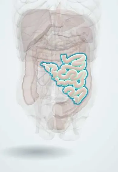
Structures involved
Tap one to see what it is and open it in 3D.
Microorganisms that cause it
Why it happens
- Health effects of climate change
- Water and electrolyte absorption and how diarrhea happens
- Dehydration and fluid overload
- Metabolic acidosis, the anion gap and compensation
- G protein-coupled receptors and cAMP
- Cyclic AMP (cAMP)
What it is
An acute secretory diarrhoeal disease caused by toxin-producing Vibrio cholerae serogroups O1 and O139. The toxin makes the small intestine pour out water and salts, producing profuse painless "rice-water" stools and rapid dehydration.
Who gets it
WHO estimates millions of cases and tens of thousands of deaths a year, concentrated where safe water and sanitation are lacking, during conflicts and after disasters. Latin America had a large epidemic that began in Peru in 1991 and reached Colombia, and Haiti has had major outbreaks since 2010. Colombia keeps cholera under mandatory surveillance; every suspected case is notified.
Causes
- Vibrio cholerae O1 (classical and El Tor biotypes) and O139, curved Gram-negative rods
- Faecal-oral spread through contaminated water, ice, street food and raw or undercooked seafood
- Asymptomatic carriers shed the bacteria in stool for days
Risk factors
- Lack of safe drinking water and sanitation
- Crowding in camps for displaced people and after disasters
- Low gastric acid (antacid use, gastrectomy)
- Blood group O (more severe disease)
- Malnutrition and young age
Pathophysiology
A large inoculum is needed because the bacteria are acid-sensitive. Survivors attach to the small-bowel mucosa without invading it and release cholera toxin. The toxin permanently activates the stimulatory G protein (Gs) by ADP-ribosylation, raising cAMP in enterocytes; chloride is pumped into the lumen and sodium absorption falls, so water follows. The colon cannot reabsorb the volume, giving isotonic watery diarrhoea that can reach litres per hour, with loss of potassium and bicarbonate. Glucose-coupled sodium absorption stays intact, which is why oral rehydration solution works.
Classification
- No dehydration
- Some dehydration
- Severe dehydration (WHO dehydration grading guides treatment)
Symptoms and signs
- Sudden, painless, profuse watery diarrhoea with a "rice-water" look and fishy smell
- Vomiting, usually without high fever
- Intense thirst, dry mouth and sunken eyes
- Reduced skin turgor and "washerwoman's hands"
- Leg cramps from electrolyte loss
- Low urine output, fast weak pulse, lethargy
Diagnosis
- Clinical
- Suspected in anyone with acute watery diarrhoea and severe dehydration in an area with cholera, or in an outbreak. Rehydration starts at once on clinical grounds; dehydration is graded at the bedside.
- Tests
- Stool culture on selective media (TCBS agar) with serogrouping, stool PCR panels and rapid diagnostic tests for outbreak detection. Electrolytes, glucose, creatinine and acid-base status in severe cases.
- Imaging
- Not needed.
Differential diagnosis
- Enterotoxigenic Escherichia coli (traveller's diarrhoea)
- Viral gastroenteritis (rotavirus, norovirus)
- Bacterial dysentery (bloody stools)
- Food poisoning from preformed toxins
- Giardiasis and other parasitic diarrhoeas
Goal of treatment
Replace lost water and salts fast enough to reverse and then prevent dehydration, keep up with ongoing losses, correct potassium and acidosis, and stop spread.
Treatment
- Acute
- Oral rehydration solution for most patients, including those with some dehydration, given in small frequent amounts and matched to ongoing stool losses.
- Acute
- Severe dehydration or shock: rapid intravenous isotonic fluid, preferably Ringer's lactate, then oral solution as soon as the patient can drink.
- Medical
- Antibiotics (for example doxycycline or azithromycin, chosen by local resistance) for moderate to severe cases shorten diarrhoea and shedding; mass prophylaxis is not recommended. Zinc for children.
- Follow-up
- Nursing priorities: frequent reassessment of pulse, mental status, thirst and urine, recording intake and output (cholera cots allow stool measurement), potassium and glucose checks; contact precautions and hand hygiene.
- Medical
- Early refeeding and continued breastfeeding to prevent malnutrition.
Complications
- Hypovolaemic shock and death within hours
- Acute kidney injury
- Hypokalaemia with muscle weakness and arrhythmia
- Metabolic acidosis from bicarbonate loss
- Hypoglycaemia in children
Prognosis
Untreated severe cholera can kill a large share of patients, but with proper rehydration mortality falls below 1%. Most people with mild infection recover without specific treatment.
Prevention
- Safe water, sanitation and hygiene (boiling or chlorinating water, hand washing)
- Food safety: cooking seafood well and avoiding raw food in outbreaks
- Oral cholera vaccines in endemic areas and outbreaks
- Rapid notification, case management centres and surveillance
Test yourself
Think of the answer, then tap the question to see it.
How does cholera toxin cause diarrhoea?
It ADP-ribosylates and permanently activates Gs, raising cAMP so enterocytes secrete chloride and water.
Why does oral rehydration solution work in cholera?
Glucose-coupled sodium absorption in the small intestine stays intact, so sodium and water are absorbed with glucose.
What does the classic cholera stool look like?
Painless, profuse "rice-water" diarrhoea, without blood, with a fishy smell.
Which fluid is preferred for severe dehydration?
Intravenous Ringer's lactate, switching to oral solution once the patient can drink.
Which electrolyte problems should the nurse watch for?
Hypokalaemia and metabolic acidosis from stool losses of potassium and bicarbonate, plus hypoglycaemia in children.
Related conditions
- Acute (viral) gastroenteritis
- Hypovolaemic shock
- Dehydration (fluid volume deficit)
- Dehydration in children (acute diarrhoeal disease)
- Acute kidney injury
- Hypokalaemia
- Bacterial infectious diarrhoea and dysentery
- Typhoid (enteric) fever
- Giardiasis
Educational information, not medical advice. For any symptom, see a health professional.
Sources: Cholera (World Health Organization, 2026); Cholera: MedlinePlus Medical Encyclopedia (MedlinePlus (U.S. National Library of Medicine), 2024).
Spotted a mistake?We are always improving for the community. Tell us what is wrong and our team will review it.Report a mistake
Representative model, for educational purposes only.