Conditions · Systemic conditions
Hypovolaemic shock !
Hypovolaemic shock is a life-threatening state in which the body loses so much blood or fluid that organs no longer get enough blood flow.
Updated 2026-10-29
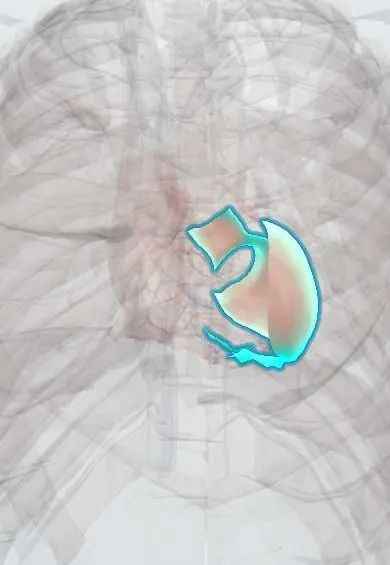
Structures involved
Tap one to see what it is and open it in 3D.
In biochemistry
In the clinical exam
- Shock: the clinical signs and the types
- Hydration status: signs of dehydration
- Capillary refill time
- Dehydration and fluid overload
In physiology
- The Frank–Starling mechanism
- Cardiac output and what sets it
- The baroreceptor reflex
- Shock physiology
- Venous return and the Guyton curves
- Central venous pressure and the jugular pulse
- Nervous control of the circulation and the CNS ischemic response
- The response to standing up
- Skin blood flow
- Body fluid compartments
- The endocrine stress response
- Water and electrolyte absorption and how diarrhea happens
- Splanchnic and portal circulation
- Physiology of the vital signs (including blood pressure measurement)
- Glomerular filtration: the barrier and its determinants
- Sodium and extracellular volume regulation (with natriuretic peptides)
- Renal blood flow, autoregulation and tubuloglomerular feedback
- Fluid shifts between compartments
- Dehydration and fluid overload
- IV fluids: tonicity and where they go
- Oxygen content, delivery and extraction
Why it happens
- Shock: types, stages and the organs it damages
- Watershed areas and the organs most sensitive to ischemia
- Thermal injury: burns, hyperthermia and hypothermia
- Hemorrhage: petechiae, purpura, ecchymosis, hematoma, and what blood loss does
What it is
A life-threatening state of poor tissue perfusion caused by loss of intravascular volume, from bleeding (haemorrhagic shock) or from fluid losses such as vomiting, diarrhoea, burns or diuresis.
Who gets it
Haemorrhage is a leading preventable cause of death after trauma, which in Colombia includes road traffic injuries and violence. Diarrhoeal dehydration remains an important cause of hypovolaemia in young children.
Causes
- Haemorrhage: trauma (chest, abdomen, pelvis, long bones, external), gastrointestinal bleeding, ruptured aortic aneurysm, ectopic pregnancy, postpartum haemorrhage
- Gastrointestinal losses: vomiting and diarrhoea
- Kidney losses: osmotic diuresis (diabetic ketoacidosis), diuretics, adrenal insufficiency
- Skin losses: burns, heat illness
- Third-space losses: pancreatitis, bowel obstruction, sepsis capillary leak
Risk factors
- Major trauma
- Anticoagulant or antiplatelet use
- Extremes of age
- Severe gastroenteritis in infants
- Limited access to early care
Pathophysiology
Reduced venous return lowers preload, stroke volume and cardiac output. Sympathetic activation causes tachycardia and vasoconstriction, keeping blood pressure normal at first. As losses grow, compensation fails: hypotension, anaerobic metabolism and lactic acidosis follow. In trauma, hypothermia, acidosis and coagulopathy reinforce each other (the lethal triad).
Classification
- Haemorrhagic shock classes (ATLS) by estimated blood loss: class I (up to about 15%), II (15 to 30%), III (30 to 40%) and IV (over 40%)
- Non-haemorrhagic hypovolaemia (fluid losses)
- Compensated or decompensated shock
Symptoms and signs
- Early: tachycardia, anxiety, cool pale skin, delayed capillary refill, narrowed pulse pressure
- Thirst, dry mucous membranes (dehydration)
- Reduced urine output
- Late: hypotension, confusion or drowsiness, rapid breathing
- Visible bleeding or signs of internal bleeding (distended abdomen, pelvic or femoral deformity)
Diagnosis
- Clinical
- A clinical diagnosis: a source of loss plus signs of hypoperfusion. In trauma the primary survey (ABCDE) looks for bleeding in the chest, abdomen, pelvis, long bones and externally; the shock index (heart rate divided by systolic pressure) helps.
- Tests
- Lactate and base deficit (perfusion), haemoglobin (may be normal early in acute bleeding), blood type and cross-match, coagulation tests or viscoelastic testing, electrolytes, kidney function and blood gas.
- Imaging
- Bedside ultrasound (FAST/eFAST) for blood in the abdomen, pericardium or chest; chest and pelvic X-rays in trauma; CT once the patient is stable; ultrasound of the inferior vena cava to estimate volume status.
Differential diagnosis
- Cardiogenic shock (myocardial infarction)
- Obstructive shock (tension pneumothorax, cardiac tamponade, pulmonary embolism)
- Septic shock
- Anaphylactic shock
- Neurogenic shock (spinal cord injury)
Goal of treatment
Stop the loss, restore circulating volume and oxygen delivery quickly, and prevent organ failure and the lethal triad of hypothermia, acidosis and coagulopathy.
Treatment
- Acute
- Haemorrhage control first: direct pressure, tourniquets, pelvic binder; urgent surgery or interventional radiology for internal bleeding.
- Acute
- Haemorrhagic shock: early balanced blood products (massive transfusion protocol), little crystalloid, permissive hypotension until bleeding is controlled (not in head injury), early antifibrinolytic in trauma.
- Acute
- Non-haemorrhagic losses: warmed isotonic crystalloids given in boluses with reassessment, and correction of electrolytes.
- Follow-up
- Heart rate, blood pressure, urine output, mental status, lactate and temperature to guide resuscitation.
- Medical
- Treating the cause: for example antiemetics, insulin and fluids in ketoacidosis, or hormone replacement in adrenal crisis.
Drugs for this condition
Complications
- Acute kidney injury
- Multiple organ dysfunction
- Coagulopathy and dilution from large volumes of crystalloid
- Hypothermia and metabolic acidosis
- Abdominal compartment syndrome
- Cardiac arrest and death
Prognosis
Depends on the cause, the amount lost and how quickly the source is controlled. Early haemorrhage control and blood product resuscitation improve survival; prolonged shock leads to irreversible organ failure.
Prevention
- Road safety and injury prevention
- Oral rehydration for diarrhoea in children
- Early recognition and treatment of bleeding in hospital and before arrival
Test yourself
Think of the answer, then tap the question to see it.
Which is an early sign of hypovolaemic shock, and which is late?
Tachycardia (with cool skin and narrowed pulse pressure) is early; hypotension is late, as compensation has failed after a large loss.
From which ATLS class of haemorrhagic shock does systolic blood pressure usually fall?
Class III (about 30 to 40% of blood volume lost); classes I and II usually keep a normal blood pressure.
What is the lethal triad of trauma?
Hypothermia, acidosis and coagulopathy, which worsen each other and increase bleeding and death.
Why is large-volume crystalloid avoided in haemorrhagic shock?
It dilutes clotting factors and red cells, cools the patient and can worsen bleeding; early balanced blood products are preferred.
Why may haemoglobin be normal right after acute bleeding?
Whole blood is lost in proportion, so the concentration falls only after fluid shifts in or is given; normal haemoglobin does not exclude major blood loss.
Related conditions
- Popliteal Artery Injury
- Postpartum haemorrhage
- Placental abruption
- Ectopic pregnancy
- Acute (viral) gastroenteritis
- Splenic injury
- Acute kidney injury
- Femoral shaft fracture
Educational information, not medical advice. For any symptom, see a health professional.
Sources: Hypovolemia and Hypovolemic Shock (StatPearls (NCBI Bookshelf), 2025).
Spotted a mistake?We are always improving for the community. Tell us what is wrong and our team will review it.Report a mistake
Representative model, for educational purposes only.