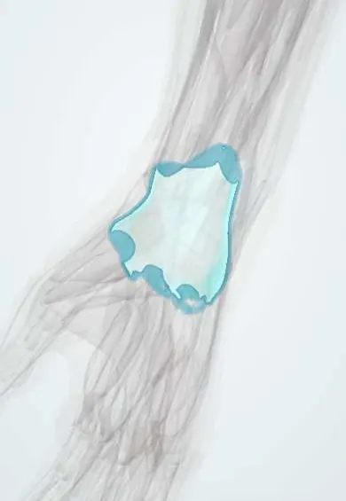
Ganglion cyst !
A ganglion cyst is a harmless fluid-filled lump from a joint or tendon sheath, most often on the back of the wrist; it is the most common hand lump.
Updated 2026-10-29
Structures involved
Tap one to see what it is and open it in 3D.
- Wrist capsule
- Scapholunate ligament
- Dorsal radiocarpal ligament
- Extensor tendon sheaths
- Flexor tendon sheaths
- Radial artery
- Scaphoid
- Lunate
Why it happens
What it is
A benign, fluid-filled swelling arising from a joint capsule or tendon sheath, most often on the back of the wrist. It is the most common soft-tissue mass of the hand and wrist.
Who gets it
Most common in women and in people between their 20s and 40s. It is a frequent reason for consultation in hand and general orthopaedic clinics.
Causes
- Usually no clear cause
- Mucoid degeneration of the joint capsule or tendon sheath
- Previous minor trauma or repetitive stress in some cases
- Underlying joint degeneration (mucous cysts at the finger DIP joints with osteoarthritis)
Risk factors
- Female sex
- Age 20 to 40
- Repetitive wrist loading (gymnasts, manual work)
- Osteoarthritis of the finger joints (mucous cysts)
Pathophysiology
A one-way valve from the joint or sheath lets synovial fluid collect in a sac without a true epithelial lining, where it thickens into a gel-like mucin. About two-thirds are dorsal and arise from the scapholunate ligament area; volar wrist ganglia usually arise near the radial artery from the radiocarpal or scaphotrapezial joint.
Classification
- Dorsal wrist ganglion (most common)
- Volar wrist ganglion
- Flexor tendon sheath (retinacular) ganglion at the base of a finger
- Mucous cyst at the distal interphalangeal joint
- Occult ganglion (not palpable)
Symptoms and signs
- Round, smooth, firm or rubbery lump that may change size
- Often painless; sometimes aching or weakness with gripping
- Pain with wrist extension, especially with small occult dorsal ganglia
- Tingling if it presses on a nearby nerve
Diagnosis
- Clinical
- Smooth, mobile, well-defined lump at a typical site. It transilluminates: a light shone through it shows a glow, unlike a solid mass. Clinicians check the nearby radial artery (Allen test for volar ganglia) and nerves.
- Tests
- Aspiration yields thick, clear, jelly-like fluid, which supports the diagnosis.
- Imaging
- Ultrasound confirms a cystic lesion and its stalk; X-ray looks for arthritis; MRI for occult ganglia or uncertain masses.
Differential diagnosis
- Lipoma
- Giant cell tumour of tendon sheath
- Tenosynovitis or rheumatoid nodule
- Carpal boss (bony bump at the base of the second and third metacarpals)
- Vascular lesion such as a radial artery aneurysm (volar)
Goal of treatment
Confirm the benign nature of the lump, relieve symptoms and reduce recurrence when treatment is needed.
Treatment
- Follow-up
- Observation for painless ganglia: many resolve on their own, and reassurance is often all that is needed.
- Procedure
- Needle aspiration, sometimes with corticosteroid injection; simple and quick but recurrence is common. Caution near the radial artery with volar ganglia.
- Surgical
- Surgical excision (open or arthroscopic) including the stalk and part of the capsule, for painful, recurrent or troublesome cysts; recurrence is lower than with aspiration but not zero.
Complications
- Recurrence after aspiration or surgery
- Wrist stiffness or scar pain after surgery
- Injury to the radial artery or nearby nerves (volar excision)
- Infection (rare)
Prognosis
Benign, with no malignant potential. Many disappear without treatment; recurrence after aspiration is common and lower after excision.
Test yourself
Think of the answer, then tap the question to see it.
What is the most common location of a wrist ganglion and from which ligament does it usually arise?
The dorsal wrist, usually from the scapholunate ligament region.
Which simple bedside test helps tell a ganglion from a solid mass?
Transillumination: a light shone through a ganglion makes it glow because it is fluid-filled.
Which artery must be considered before aspirating a volar wrist ganglion?
The radial artery, which often lies very close to volar ganglia.
Does a ganglion cyst have a true epithelial lining?
No. Its wall is compressed connective tissue without a true synovial or epithelial lining, which is why it is not a true cyst.
Related conditions
Educational information, not medical advice. For any symptom, see a health professional.
Sources: Ganglion Cyst (StatPearls (NCBI Bookshelf), 2023).
Spotted a mistake?We are always improving for the community. Tell us what is wrong and our team will review it.Report a mistake
Representative model, for educational purposes only.