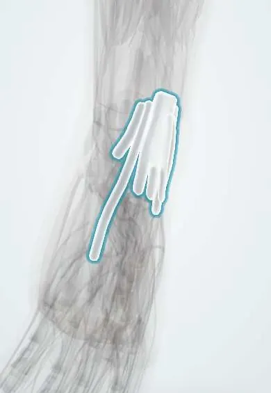
Extensor tendon sheaths !
The extensor tendon sheaths are fluid-lined tubes on the back of the wrist; they let the extensor tendons glide under the extensor retinaculum.
Official name: Wrist extensor tendon sheaths
Opens in the Durgical app, no sign-up needed.
What it is
Sleeves around the extensor tendons in the tunnels on the back of the wrist.
What it does
Let the extensor tendons glide under the retinaculum.
Key facts
- Where
- Six dorsal compartments under the extensor retinaculum: 1 APL/EPB; 2 ECRL/ECRB; 3 EPL; 4 ED/EIP; 5 EDM; 6 ECU
- Function
- Let the extensor tendons glide under the retinaculum
Relations
- Contents
- Six synovial sheaths, one per compartment; the extensor digitorum and extensor indicis share the 4th
- Superficial
- Extensor retinaculum; over it the superficial radial nerve, the dorsal branch of the ulnar nerve and the origins of the cephalic and basilic veins
- Deep
- Grooves of the distal radius and ulna and the dorsal wrist capsule; the posterior interosseous nerve lies in the floor of the 4th compartment
- Below
- The sheaths end over the carpus and bases of the metacarpals; the tendons then run under the dorsal hand fascia
How to find it
Lister's tubercle is felt on the back of the distal radius, roughly in line with the third metacarpal; the EPL runs on its ulnar side, the ECRL and ECRB on its radial side. The ECU is felt in its groove beside the ulnar head.
Examination
- Thumb lift test: with the palm flat, inability to lift the thumb off the table suggests EPL rupture
- ECU synergy test: resisted thumb abduction with the forearm supinated reproduces ulnar-sided wrist pain in ECU tendinitis
On imaging
Ultrasound shows fluid in a sheath, ECU subluxation on dynamic scanning and the gap of a ruptured tendon; MRI shows tenosynovitis in inflammatory arthritis.
Clinical notes
- EPL rupture is usually treated by transferring the extensor indicis tendon
- Dorsal wrist arthroscopy portals are named after the compartments: 3-4, 4-5, 6R and 6U
- Rheumatoid dorsal tenosynovitis may need tenosynovectomy to prevent tendon rupture
Anatomical variants
- A septum often splits the 1st compartment, giving the EPB its own tunnel
- The extensor digiti minimi in the 5th compartment is commonly doubled, and extra slips of the extensor indicis and extensor digitorum are frequent
Memory aid
Tendons per compartment, from the radial side: 2-2-1-2-1-1 (APL/EPB, ECRL/ECRB, EPL, ED/EIP, EDM, ECU).
Related conditions
Test yourself
Think of the answer, then tap the question to see it.
Which tendon runs in the 3rd extensor compartment, and why can it rupture after a distal radius fracture?
The extensor pollicis longus (EPL); it turns around Lister's tubercle and can rupture there weeks later, often after an undisplaced fracture.
What is the Vaughan-Jackson lesion?
In rheumatoid arthritis, extensor tendons rupture from the ulnar side first over an eroded ulnar head.
Which test suggests extensor pollicis longus rupture?
The thumb lift test: with the palm flat, the thumb cannot be lifted off the table.
Which tendon has its own subsheath in the 6th extensor compartment, and how does it fail?
The extensor carpi ulnaris (ECU), in a groove of the ulna; it can sublux with supination and ulnar deviation.
Educational information, not medical advice. For any symptom, see a health professional.
Its names
- Official Latin (TA2)
- vagina tendinis extensoris ulnaris carpi TA2 2582
- Official Spanish (DeCS)
- Vainas tendinosas carpianas dorsales
- English (TA2)
- Wrist extensor tendon sheaths
Related
Sources: Terminologia Anatomica (TA2); Anatomy, Shoulder and Upper Limb, Wrist Extensor Muscles.
Spotted a mistake?We are always improving for the community. Tell us what is wrong and our team will review it.Report a mistake
Representative model, for educational purposes only.