Conditions · Pediatrics and neonatology
Gastroschisis and omphalocele !
Two birth defects of the belly wall where bowel lies outside the body: gastroschisis is uncovered, omphalocele is covered by a sac. Both need surgery.
Updated 2026-11-01
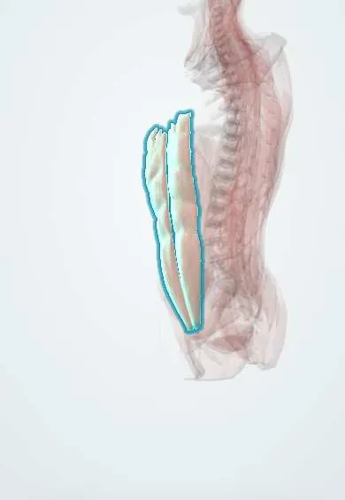
Structures involved
Tap one to see what it is and open it in 3D.
What it is
Congenital abdominal wall defects in which abdominal contents lie outside the body at birth. In gastroschisis the bowel herniates through a full-thickness defect beside the umbilical cord, usually to its right, with no covering membrane. In omphalocele the viscera herniate through the umbilical ring itself and are covered by a membranous sac (amnion and peritoneum) with the cord inserting into it.
Who gets it
Each affects roughly 1 in a few thousand births. Gastroschisis has risen worldwide and is linked to young maternal age; omphalocele is more often linked to older maternal age and to chromosomal syndromes. In Colombia and Latin America both are detected more often now by routine prenatal ultrasound, and outcomes depend heavily on delivery in a centre with neonatal surgery and intensive care.
Causes
- Gastroschisis: thought to arise from a vascular disruption of the right paraumbilical body wall in early pregnancy; usually an isolated defect
- Omphalocele: failure of the midgut to return into the abdomen after physiological herniation (around weeks 10–12), with failed closure of the umbilical ring
- Omphalocele is often part of a syndrome: trisomies 13, 18 and 21, Beckwith–Wiedemann syndrome, pentalogy of Cantrell
- Gastroschisis is associated with intestinal atresia in a minority of cases
Risk factors
- Gastroschisis: maternal age under 20 years
- Gastroschisis: maternal smoking, alcohol, recreational drugs and vasoactive medicines in early pregnancy
- Omphalocele: advanced maternal age
- Omphalocele: chromosomal anomalies and overgrowth syndromes
- Low socioeconomic status and poor periconceptional nutrition (gastroschisis)
Pathophysiology
In gastroschisis the exposed bowel floats in amniotic fluid without a covering, so it becomes thickened, matted and inflamed (a fibrinous peel), with poor motility after birth; the defect is small, which can strangle the bowel. In omphalocele the sac protects the viscera, but large defects may contain liver and leave a small abdominal cavity and underdeveloped lungs. After birth both lose heat and water rapidly from the exposed surface, and forcing the viscera back into a small abdomen can raise intra-abdominal pressure and cause abdominal compartment syndrome.
Classification
- Gastroschisis: simple (bowel intact) or complex (with atresia, perforation, necrosis or volvulus)
- Omphalocele: minor (small, bowel only) or giant (large defect, usually containing liver)
- Isolated or syndromic (with chromosomal, cardiac or other anomalies), mainly relevant to omphalocele
Symptoms and signs
- Bowel visible outside the abdomen at birth
- Gastroschisis: uncovered, often thickened bowel to the right of a normally inserted cord
- Omphalocele: central bulge covered by a translucent sac, with the cord arising from the sac
- Omphalocele may contain liver, stomach or spleen
- Associated features in omphalocele: macroglossia and hypoglycaemia (Beckwith–Wiedemann), heart defects
Diagnosis
- Clinical
- Usually diagnosed on prenatal ultrasound in the second trimester and confirmed at birth by inspection: location of the defect relative to the cord and presence or absence of a sac. Raised maternal serum alpha-fetoprotein is a screening clue for both.
- Tests
- For omphalocele: fetal karyotype or chromosomal microarray and, after birth, glucose monitoring. For both: blood count, electrolytes, glucose and blood gases in the newborn.
- Imaging
- Prenatal ultrasound with serial growth and bowel assessment; fetal echocardiography (especially for omphalocele); postnatal echocardiography and renal ultrasound to look for associated anomalies.
Differential diagnosis
- Ruptured omphalocele (can mimic gastroschisis, but has sac remnants and a central defect)
- Umbilical cord hernia
- Bladder or cloacal exstrophy
- Pentalogy of Cantrell or limb–body wall complex
- Umbilical hernia (skin-covered, benign)
Goal of treatment
Protect the exposed bowel, prevent heat and fluid loss and infection, return the viscera to the abdomen without raising abdominal pressure dangerously, and support nutrition until the gut works.
Treatment
- Acute
- Delivery room: wrap or bag the exposed bowel in sterile, moist, clear covering, keep the infant warm, lay them on the side to avoid kinking the mesentery, place a nasogastric tube to decompress, start intravenous fluids and broad-spectrum antibiotics.
- Surgical
- Gastroschisis: primary closure when the bowel reduces easily, or staged reduction in a preformed silo with gradual return of the bowel over days, then closure of the defect.
- Surgical
- Omphalocele: early repair for small defects; giant defects may be managed with a staged repair or with topical treatment of the sac to let it epithelialise, and delayed closure of the ventral hernia later.
- Medical
- Parenteral nutrition until bowel function returns, then gradual enteral feeding, ideally with breast milk.
- Follow-up
- Nursing monitoring: temperature, fluid balance and urine output, bowel colour, bladder or abdominal pressure and breathing after reduction, glucose in omphalocele, signs of infection.
Complications
- Prolonged intestinal dysmotility and feeding intolerance (gastroschisis)
- Intestinal atresia, necrosis or volvulus
- Short bowel syndrome and parenteral-nutrition liver disease
- Abdominal compartment syndrome after closure
- Sepsis and central line infections
- Pulmonary hypoplasia (giant omphalocele)
- Necrotising enterocolitis
Prognosis
Most infants with simple gastroschisis survive with good long-term function, though hospital stays can be long. In omphalocele the outlook depends mostly on the associated chromosomal and heart anomalies and on lung development in giant defects.
Prevention
- Avoidance of tobacco, alcohol and drugs in pregnancy
- Prenatal ultrasound to plan delivery in a centre with neonatal surgery
- Folic acid and good nutrition before and during pregnancy
Test yourself
Think of the answer, then tap the question to see it.
Which abdominal wall defect has a covering sac and is linked to trisomies and Beckwith–Wiedemann syndrome?
Omphalocele.
Where is the defect in gastroschisis relative to the umbilical cord?
Usually to the right of a normally inserted cord, with no covering sac.
What maternal factor is classically associated with gastroschisis?
Young maternal age (under 20 years).
What is the first priority in the delivery room for exposed bowel?
Cover it with a sterile moist clear wrap, keep the baby warm and decompress the stomach.
Which prenatal screening marker is raised in both defects?
Maternal serum alpha-fetoprotein.
Related conditions
- Duodenal and intestinal atresia
- Short bowel syndrome
- Umbilical hernia
- Beckwith–Wiedemann syndrome
- Congenital diaphragmatic hernia
- Sepsis and septic shock
Educational information, not medical advice. For any symptom, see a health professional.
Sources: Gastroschisis | Birth Defects | CDC (Centers for Disease Control and Prevention, 2024); Omphalocele: MedlinePlus Medical Encyclopedia (MedlinePlus (U.S. National Library of Medicine), 2024).
Spotted a mistake?We are always improving for the community. Tell us what is wrong and our team will review it.Report a mistake
Representative model, for educational purposes only.