Conditions · Gastroenterology and hepatology
Short bowel syndrome !
Malabsorption after too much small bowel is removed or lost. It causes diarrhoea, dehydration and malnutrition; some patients need IV feeding.
Updated 2026-11-01
Structures involved
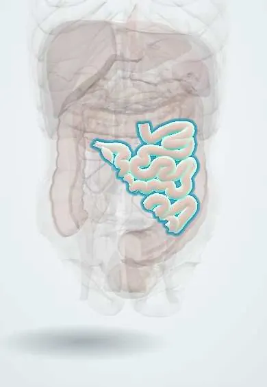
Tap one to see what it is and open it in 3D.
Why it happens
- Necrotizing enterocolitis: why the premature gut is vulnerable
- Patterns of malabsorption
- Bile and the enterohepatic circulation
- Absorption of iron, calcium and vitamin B12
- Water and electrolyte absorption and how diarrhea happens
- Small intestine: villi, crypts, enterocytes, Paneth cells; duodenum vs jejunum vs ileum
What it is
A malabsorption state that results from loss of a large length of functioning small bowel, usually after surgical resection. The remaining bowel cannot absorb enough fluid, electrolytes and nutrients, so patients develop diarrhoea, dehydration and malnutrition, and may depend on parenteral nutrition (intestinal failure).
Who gets it
Uncommon but a leading cause of intestinal failure in adults and children. In adults it follows extensive resection for mesenteric ischaemia, Crohn disease, trauma, volvulus or radiation injury; in infants, necrotising enterocolitis and congenital anomalies are the main causes. Care depends on specialised nutrition teams that are concentrated in large hospitals in Colombia.
Causes
- Extensive small bowel resection, usually leaving less than about 200 cm
- Adults: acute mesenteric ischaemia, Crohn disease, trauma, volvulus, radiation enteritis, cancer surgery
- Children: necrotising enterocolitis, midgut volvulus with malrotation, intestinal atresia, gastroschisis
- Functional loss without resection (extensive mucosal disease) is less common
Risk factors
- Repeated bowel surgery (Crohn disease)
- Atherosclerosis and atrial fibrillation (mesenteric ischaemia)
- Prematurity (necrotising enterocolitis)
- Abdominal or pelvic radiotherapy
Pathophysiology
Outcome depends on the length and site of bowel remaining and whether the colon and ileocaecal valve are kept. The jejunum absorbs most nutrients but cannot adapt to take over ileal functions; the terminal ileum absorbs vitamin B12 and bile acids. Losing the ileum leads to B12 deficiency, bile acid diarrhoea and, with larger losses, fat malabsorption and fat-soluble vitamin deficiency. Unabsorbed fat binds calcium in the gut, so oxalate is absorbed freely in the colon and forms kidney stones; bile salt loss promotes gallstones. Gastric acid hypersecretion after resection inactivates pancreatic enzymes, and loss of the ileocaecal valve allows bacterial overgrowth. Over months, the remaining bowel adapts by growing longer villi.
Classification
- End jejunostomy (no colon in continuity): highest fluid losses
- Jejunocolic anastomosis (part of colon kept)
- Jejunoileal anastomosis with ileocaecal valve and colon kept: best prognosis
Symptoms and signs
- High-volume diarrhoea or high stoma output
- Dehydration, thirst, low urine output
- Weight loss and muscle wasting
- Fatty, foul-smelling stools
- Fatigue and weakness
- Signs of deficiency: anaemia, bruising, bone pain, night blindness, tetany
Diagnosis
- Clinical
- Based on the surgical history (length and site of bowel removed, colon kept or not) and the clinical picture of malabsorption and high fluid losses.
- Tests
- Regular monitoring of electrolytes (sodium, potassium, magnesium, calcium, phosphate), kidney and liver function, blood count, albumin, vitamin B12, fat-soluble vitamins, zinc and iron. Urine sodium helps assess sodium depletion. Stool or stoma output is measured.
- Imaging
- Contrast studies or CT/MR enterography estimate remaining bowel length and detect strictures; bone densitometry for metabolic bone disease; ultrasound for gallstones and kidney stones.
Differential diagnosis
- Small intestinal bacterial overgrowth
- Recurrent Crohn disease
- Infectious diarrhoea including Clostridioides difficile
- Bile acid diarrhoea
- Pancreatic exocrine insufficiency
Goal of treatment
Keep the patient hydrated and nourished, promote intestinal adaptation, prevent complications and reduce or stop dependence on parenteral nutrition.
Treatment
- Acute
- Early after resection: intravenous fluids, electrolytes and parenteral nutrition, with strict fluid balance.
- Lifestyle
- Gradual oral or enteral feeding with small, frequent meals; oral rehydration solutions rich in sodium instead of plain water or sweet drinks; in patients with a colon, a low-oxalate diet.
- Medical
- Antidiarrhoeal agents before meals, proton pump inhibitors or H2 blockers for acid hypersecretion, bile acid sequestrants for bile acid diarrhoea (when the colon is kept and ileal loss is limited), vitamin B12 by injection and other vitamin and mineral replacement. GLP-2 analogues (teduglutide) increase adaptation in patients dependent on parenteral support.
- Surgical
- Intestinal lengthening procedures (such as serial transverse enteroplasty) or intestinal transplantation for patients who fail parenteral nutrition.
- Follow-up
- Nursing care of the central venous catheter with strict aseptic technique, daily weight, intake and output, stoma output, blood glucose during parenteral nutrition and periodic laboratory tests.
Complications
- Dehydration, acute kidney injury and electrolyte disturbances (low magnesium, potassium, calcium)
- Central venous catheter infection and thrombosis
- Intestinal failure-associated liver disease (cholestasis, fibrosis)
- Calcium oxalate kidney stones
- Gallstones
- Vitamin B12 and fat-soluble vitamin deficiencies; metabolic bone disease
- D-lactic acidosis and bacterial overgrowth
Prognosis
Depends on remaining bowel length, presence of the colon and ileocaecal valve, and the underlying disease. Many patients wean from parenteral nutrition as the bowel adapts over 1 to 2 years; those with very short bowel (less than about 100 cm without colon) usually need long-term parenteral support. Children have a greater capacity for adaptation.
Prevention
- Bowel-sparing surgery when possible (in Crohn disease, ischaemia and trauma)
- Early recognition and treatment of mesenteric ischaemia and volvulus
- Prevention of necrotising enterocolitis in premature infants (breast milk feeding)
Test yourself
Think of the answer, then tap the question to see it.
Which part of the small bowel absorbs vitamin B12 and bile acids?
The terminal ileum.
Why do patients with short bowel and an intact colon form kidney stones?
Unabsorbed fat binds calcium, so free oxalate is absorbed in the colon (enteric hyperoxaluria) and forms calcium oxalate stones.
What is the most common cause in infants?
Necrotising enterocolitis (also midgut volvulus and intestinal atresia).
What is the main life-threatening complication of parenteral nutrition in these patients?
Central venous catheter-related bloodstream infection (sepsis); liver disease is another.
Which hormone analogue promotes intestinal adaptation?
GLP-2 analogues such as teduglutide.
Related conditions
- Crohn disease
- Acute mesenteric ischaemia
- Necrotising enterocolitis
- Intestinal malrotation with midgut volvulus
- Duodenal and intestinal atresia
- Small intestinal bacterial overgrowth (SIBO)
- Kidney stones (urolithiasis)
- Gallstones (cholelithiasis)
- Megaloblastic anaemia (B12 or folate deficiency)
- Osteoporosis
Educational information, not medical advice. For any symptom, see a health professional.
Sources: Short Bowel Syndrome - Gastroenterology - MSD Manual Professional Edition (MSD Manual Professional Edition, 2024); Short Bowel Syndrome (National Institute of Diabetes and Digestive and Kidney Diseases (NIDDK), 2025).
Spotted a mistake?We are always improving for the community. Tell us what is wrong and our team will review it.Report a mistake
Representative model, for educational purposes only.