Conditions · Pediatrics and neonatology
Congenital diaphragmatic hernia !
A hole in the diaphragm lets abdominal organs move into the chest before birth, so the lungs grow too small. Babies need intensive care and surgery.
Updated 2026-11-01
Structures involved
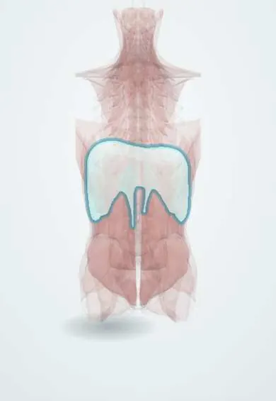
Tap one to see what it is and open it in 3D.
Why it happens
What it is
A developmental defect of the diaphragm through which abdominal organs herniate into the chest during fetal life, causing pulmonary hypoplasia and pulmonary hypertension. It is usually on the left side, through the posterolateral (Bochdalek) foramen.
Who gets it
About 1 in 2,500 to 4,000 births. Many cases are detected on routine prenatal ultrasound. In Latin America, survival depends heavily on delivery in a centre with neonatal intensive care, ventilation and paediatric surgery.
Causes
- Failure of the pleuroperitoneal membranes to close in early gestation
- Usually sporadic; sometimes part of chromosomal anomalies or syndromes
Risk factors
- Associated chromosomal anomalies (e.g. trisomy 18) and genetic syndromes
- Other congenital defects, especially cardiac
Pathophysiology
Bowel, stomach, spleen and sometimes liver occupy the chest while the lungs are developing. The ipsilateral lung (and to a lesser degree the other lung) is hypoplastic, with fewer airways and alveoli and abnormally muscular pulmonary arteries. After birth this causes respiratory failure and persistent pulmonary hypertension with right-to-left shunting.
Classification
- Bochdalek (posterolateral), most cases, usually left-sided
- Morgagni (anterior, retrosternal), rarer and often milder
- Left-sided versus right-sided (right-sided often with liver herniation)
- Isolated versus associated with other anomalies
Symptoms and signs
- Severe respiratory distress and cyanosis at or soon after birth
- Scaphoid (sunken) abdomen
- Barrel-shaped chest with decreased breath sounds on the affected side
- Bowel sounds heard in the chest
- Heart sounds displaced to the right (in left-sided hernia)
Diagnosis
- Clinical
- Often known from prenatal ultrasound. At birth: respiratory distress, scaphoid abdomen and displaced heart sounds.
- Tests
- Blood gases and pre- and post-ductal pulse oximetry; karyotype or microarray; screening for associated anomalies.
- Imaging
- Prenatal ultrasound and fetal MRI (lung-to-head ratio, liver position, lung volume) predict severity. Postnatal chest X-ray shows bowel loops in the chest, mediastinal shift and the gastric tube tip above the diaphragm. Echocardiography assesses pulmonary hypertension and heart defects.
Differential diagnosis
- Congenital pulmonary airway malformation
- Eventration of the diaphragm
- Bronchopulmonary sequestration
- Pneumothorax or other causes of neonatal respiratory failure
Goal of treatment
Stabilise breathing and circulation with gentle ventilation, control pulmonary hypertension, and repair the diaphragm once the baby is physiologically stable.
Treatment
- Acute
- Intubation immediately at birth (avoid bag-mask ventilation), nasogastric tube to decompress the bowel, and gentle ventilation that limits lung injury.
- Medical
- Pulmonary vasodilators such as inhaled nitric oxide, inotropes and fluids to support circulation, and sedation.
- Procedure
- ECMO for refractory respiratory failure in centres that offer it; fetal endoscopic tracheal occlusion in selected severe cases in specialised centres.
- Surgical
- Delayed surgical repair of the diaphragm (primary closure or patch) once stable.
- Follow-up
- Long-term follow-up of lung function, pulmonary hypertension, nutrition, reflux, hearing and neurodevelopment.
Complications
- Pulmonary hypoplasia and persistent pulmonary hypertension
- Pneumothorax
- Chronic lung disease
- Gastro-oesophageal reflux and poor growth
- Recurrence of the hernia, especially with patch repair
- Chest wall deformities and scoliosis
Prognosis
Survival in experienced centres is around 70 to 90% for isolated cases and lower with associated anomalies, liver herniation or severe pulmonary hypoplasia. Survivors often have long-term respiratory and feeding problems.
Test yourself
Think of the answer, then tap the question to see it.
Which type and side of congenital diaphragmatic hernia is most common?
The posterolateral Bochdalek hernia, usually on the left side.
What are the two main causes of death in congenital diaphragmatic hernia?
Pulmonary hypoplasia and persistent pulmonary hypertension of the newborn.
What is the classic abdominal finding in a newborn with congenital diaphragmatic hernia?
A scaphoid (sunken) abdomen, because the bowel is in the chest.
Why is bag-mask ventilation avoided at birth in congenital diaphragmatic hernia?
It inflates the bowel in the chest and further compresses the lungs; immediate intubation is preferred.
Is diaphragm repair an emergency right after birth?
No. Surgery is delayed until breathing and pulmonary hypertension are stabilised.
Related conditions
- Persistent pulmonary hypertension of the newborn
- Pneumothorax
- Gastro-oesophageal reflux disease (GERD)
- Hiatal hernia
- Bronchopulmonary dysplasia
- Failure to thrive (faltering growth)
Educational information, not medical advice. For any symptom, see a health professional.
Sources: Diaphragmatic hernia (MedlinePlus (U.S. National Library of Medicine), 2024).
Spotted a mistake?We are always improving for the community. Tell us what is wrong and our team will review it.Report a mistake
Representative model, for educational purposes only.