Conditions · Metabolism (biochemistry)
Hurler syndrome (mucopolysaccharidosis type I) !
In Hurler syndrome complex sugars build up almost everywhere: coarse facial features, cloudy corneas, misshapen bones, and heart and brain disease.
Updated 2026-11-01
Structures involved
Tap one to see what it is and open it in 3D.
- Cornea
- Liver
- Spleen
- Mitral anterior leaflet
- Aortic right coronary cusp
- T12 vertebra
- L1 vertebra
- Tongue
- Carpal tunnel
In biochemistry
- Lysosomal storage diseases
- Glycosaminoglycans (mucopolysaccharides)
- Mannose 6-phosphate tagging (sending enzymes to the lysosome)
- Hyaluronic acid (hyaluronan)
- α-L-Iduronidase
- Glycoproteins and protein glycosylation (N- and O-linked)
- Iduronate 2-sulfatase (I2S)
Why it happens
What it is
An autosomal recessive mucopolysaccharidosis caused by α-L-iduronidase deficiency (IDUA gene); dermatan sulfate and heparan sulfate accumulate. "Hurler" is the severe form of MPS I.
Who gets it
Severe MPS I occurs in about 1 in 100,000 newborns; the attenuated form in about 1 in 500,000.
Causes
- Biallelic variants of the IDUA gene (autosomal recessive)
Risk factors
- Carrier parents or an affected sibling
- Consanguinity
Pathophysiology
Without α-L-iduronidase, dermatan sulfate and heparan sulfate cannot be broken down past the terminal iduronic acid and fill the lysosomes of almost all tissues. Storage thickens skin, tongue, airways, heart valves and cornea; disturbs bone growth (dysostosis multiplex) and joints; enlarges liver and spleen; and heparan sulfate stored in neurons causes cognitive decline.
Classification
- Severe MPS I (Hurler): onset in the first year, with cognitive decline
- Attenuated MPS I (Hurler–Scheie and Scheie): later onset, normal or near-normal intelligence
Symptoms and signs
- Coarse facial features appearing in the first or second year
- Large tongue and noisy breathing
- Cloudy corneas
- Large abdomen with umbilical and inguinal hernias
- A hump at the thoracolumbar junction and stiff joints (claw hands)
- Frequent ear infections and hearing loss
- Developmental delay that later regresses
Diagnosis
- Clinical
- An infant or toddler with coarse features, corneal clouding, hepatosplenomegaly, hernias, gibbus and joint stiffness.
- Tests
- High urinary glycosaminoglycans (dermatan and heparan sulfate); low α-L-iduronidase activity in leukocytes or dried blood spots; IDUA gene testing; audiometry.
- Imaging
- X-rays with dysostosis multiplex (oar-shaped ribs, beaked vertebrae, J-shaped sella, pointed metacarpals); echocardiogram with thickened mitral and aortic valves; brain and cervical spine MRI.
Treatment
- Procedure
- Haematopoietic stem cell transplant before age 2 to 2.5: preserves cognition and is the treatment of choice in the severe form.
- Medical
- Intravenous enzyme replacement with recombinant α-L-iduronidase, before and around transplant and in attenuated forms.
- Surgical
- Adenotonsillectomy, hernia repair, carpal tunnel release, spinal surgery, shunting for hydrocephalus and valve replacement as needed.
- Follow-up
- Cardiac, respiratory, hearing, eye, orthopaedic and neurodevelopmental follow-up.
Complications
- Airway obstruction and sleep apnoea
- Valve disease and heart failure
- Communicating hydrocephalus
- Cervical cord compression
- Carpal tunnel syndrome
- Loss of vision and hearing
Prognosis
Untreated, the severe form is usually fatal in the first decade from heart or breathing complications. Early transplant improves survival and cognition, though bone problems persist.
Test yourself
Think of the answer, then tap the question to see it.
Which enzyme is missing in Hurler syndrome, and what builds up?
α-L-Iduronidase; dermatan sulfate and heparan sulfate accumulate.
Which eye sign separates Hurler from Hunter syndrome?
Corneal clouding: present in Hurler, absent in Hunter.
Why is stem cell transplant preferred before age 2 in severe Hurler syndrome?
Donor cells bring enzyme to the brain and preserve cognition if transplanted before decline.
What is the set of bone changes in the mucopolysaccharidoses called?
Dysostosis multiplex.
Related conditions
- Hunter syndrome (mucopolysaccharidosis type II)
- I-cell disease (mucolipidosis II)
- Carpal tunnel syndrome
- Obstructive sleep apnoea
- Hydrocephalus
- Umbilical hernia
- Inguinal hernia
Educational information, not medical advice. For any symptom, see a health professional.
Sources: Mucopolysaccharidosis type I: MedlinePlus Genetics (MedlinePlus, US National Library of Medicine, 2026); IDUA gene: MedlinePlus Genetics (MedlinePlus, US National Library of Medicine, 2026); Textbook of Biochemistry for Medical Students, 6th ed. (Jaypee Brothers Medical Publishers (Vasudevan DM, Sreekumari S, Vaidyanathan K), 2011).
Spotted a mistake?We are always improving for the community. Tell us what is wrong and our team will review it.Report a mistake
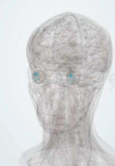
Representative model, for educational purposes only.