
Idiopathic orbital inflammation
Painful inflammation of the orbit without infection or another known cause. It responds fast to corticosteroids, but other causes must be excluded first.
What it is
A non-infectious inflammation of orbital muscles, lacrimal gland, fat or optic nerve surroundings with no identifiable cause. It causes sudden painful swelling and proptosis, responds dramatically to corticosteroids, and is diagnosed only after excluding infection, thyroid eye disease, vasculitis, IgG4-related disease and lymphoma.
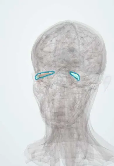
See it in 3D
Who gets it
Affects adults of any age, usually one side; less common in children, in whom it can be bilateral.
Causes
- Unknown (immune-mediated)
- Some cases are later found to be IgG4-related disease, sarcoidosis or vasculitis
Risk factors
- Autoimmune disease
- Previous episodes (recurrences are common)
Pathophysiology
A polymorphous inflammatory infiltrate with variable fibrosis involves one or more orbital structures. Muscle involvement (myositis) causes pain on movement and diplopia; lacrimal involvement (dacryoadenitis) swells the outer upper lid; apex involvement can compress the optic nerve.
Classification
- By site: myositis, dacryoadenitis, anterior, diffuse, apical
- Acute, subacute or chronic sclerosing
Symptoms and signs
- Sudden orbital pain, worse with eye movement
- Lid swelling and redness
- Proptosis
- Double vision
- Reduced vision when the apex is involved
Diagnosis
- Clinical
- Clinical picture of acute painful orbital inflammation with normal temperature and no sinus infection.
- Tests
- Thyroid tests, blood count, inflammatory markers, ANCA, ACE, IgG4; biopsy if atypical, recurrent or not responding.
- Imaging
- Contrast CT or MRI of the orbits: enhancement of a muscle including its tendon, the lacrimal gland or diffuse orbital tissue.
Differential diagnosis
- Orbital cellulitis
- Thyroid eye disease (painless, spares tendons)
- Lymphoma
- Granulomatosis with polyangiitis
- IgG4-related disease
Painful or swollen orbit: which one?
| Feature | Idiopathic orbital inflammation | Orbital cellulitis | Thyroid eye disease | Orbital lymphoma |
|---|---|---|---|---|
| Onset | Acute, hours to days | Acute, often after sinusitis | Gradual, over months | Slow, over months |
| Pain | Marked, worse on eye movement | Marked, with fever and malaise | Mild ache or grittiness | Usually painless |
| Imaging | Enlarged muscle including the tendon, swollen lacrimal gland or diffuse enhancement | Sinus opacity, fat stranding, possible subperiosteal abscess | Enlarged muscle bellies with tendons spared | Mass moulding around orbital structures |
| Sides | Usually one | One | Usually both | One or both |
| Response to corticosteroids | Rapid and dramatic | Worsens without antibiotics | Partial, only in the active phase | May shrink briefly, then recurs |
Goal of treatment
Control inflammation quickly to relieve pain and protect vision, after excluding infection and tumour.
Treatment
- Medical
- Systemic corticosteroids with a slow taper under specialist supervision.
- Medical
- Steroid-sparing immunosuppressants or biologic drugs for recurrent or resistant disease.
- Procedure
- Radiotherapy in selected resistant cases.
Complications
- Recurrence
- Compressive optic neuropathy
- Restrictive strabismus and chronic fibrosis
Common traps
- Rapid improvement on corticosteroids confirms the diagnosis: lymphoma and other mimics can also shrink on steroids, so a partial or short-lived response does not exclude them; atypical or recurrent cases are biopsied
- A painful swollen orbit without fever cannot be cellulitis: orbital cellulitis can occur without high fever, so look for sinusitis on CT and exclude infection before giving steroids
- Enlarged extraocular muscles always mean thyroid eye disease: orbital myositis enlarges the muscle including its tendon and is painful; thyroid eye disease typically spares the tendon and is usually painless
- Orbital pseudotumour is a true neoplasm: it is an inflammatory infiltrate with variable fibrosis; the old name refers only to its mass effect
- Starting corticosteroids before any work-up does no harm: steroids can mask lymphoma on a later biopsy and let an untreated infection spread, so image and exclude infection first
Prognosis
Most respond quickly; recurrences are common and sclerosing forms respond poorly.
Test yourself
Think of the answer, then tap the question to see it.
A 40-year-old has two days of painful left proptosis worse on eye movement, no fever, and CT shows a thick medial rectus including its tendon. Most likely diagnosis?
Idiopathic orbital inflammation (orbital myositis): acute pain and tendon involvement favour it over thyroid eye disease, once infection is excluded.
Why must orbital cellulitis be excluded before starting corticosteroids?
Steroids suppress the immune response; given to an untreated orbital infection they can let it spread to the cavernous sinus or brain.
A patient's orbital inflammation recurs every time the steroids are tapered. What should be done?
Biopsy, to look for lymphoma, IgG4-related disease, sarcoidosis or vasculitis; then consider steroid-sparing immunosuppression.
Why does inflammation at the orbital apex threaten vision?
The optic nerve passes through a tight bony space at the apex, so swelling there compresses it, causing visual loss and an afferent pupillary defect.
Why it happens
Connections
Educational information, not medical advice. For any symptom, see a health professional.
Sources: Nonspecific Orbital Inflammation (StatPearls Publishing (NCBI Bookshelf), 2024).
Spotted a mistake?We are always improving for the community. Tell us what is wrong and our team will review it.Report a mistake
Representative model, for educational purposes only.