Malignant pleural mesothelioma !
An aggressive cancer of the lining around the lungs, almost always linked to asbestos exposure decades earlier.
Updated 2026-11-01
Structures involved
Tap one to see what it is and open it in 3D.
Why it happens
- Air pollution and occupational exposures
- Tumour suppressor genes: RB, TP53, APC, BRCA and the two-hit hypothesis
- Chemical carcinogens: initiation, promotion and the table to know
- Naming tumours: -oma, carcinoma, sarcoma, and the misleading names
- Cytology: the Pap test, fine-needle aspiration and body fluids
- Immunohistochemistry: the principle and the markers to know
Show 1 more
What it is
A malignant tumour arising from the mesothelial cells of the pleura. It spreads along the pleural surfaces, encases the lung in a thick rind and usually presents with a recurrent pleural effusion. The peritoneum, pericardium and tunica vaginalis can also be the site of origin.
Who gets it
Rare; most patients are men over 60 with past occupational asbestos exposure. The latency between first exposure and diagnosis is typically 30 to 50 years, so cases keep appearing long after exposure ends. Colombia banned all types of asbestos by law in 2019, with a transition until 2021; asbestos-cement roofing, water tanks, brake linings and older buildings mean that exposed workers and their families remain at risk for decades.
Causes
- Asbestos exposure, especially amphibole fibres (crocidolite, amosite); chrysotile also carries risk
- Occupational settings: mining, asbestos-cement manufacturing, construction and demolition, shipbuilding, brake and clutch repair
- Para-occupational and environmental exposure: household contacts washing work clothes, living near mines or factories
- Prior chest radiation therapy
- Germline BAP1 mutations in a minority of familial cases
Risk factors
- Cumulative asbestos dose and long time since first exposure
- Male sex and older age (reflecting occupational history)
- Household members of asbestos workers
- Previous radiotherapy to the chest
- Smoking does not cause mesothelioma but multiplies the asbestos-related risk of lung cancer
Pathophysiology
Inhaled asbestos fibres are long and thin, resist clearance and migrate to the pleura. Macrophages that fail to engulf them release reactive oxygen species and inflammatory cytokines; chronic inflammation and direct fibre damage to chromosomes lead to loss of tumour suppressor genes such as BAP1, CDKN2A and NF2. The tumour grows as nodules that coalesce into a sheet over the parietal and visceral pleura, invading the chest wall, diaphragm, pericardium and mediastinum.
Classification
- Epithelioid (most common, best prognosis)
- Sarcomatoid (worst prognosis)
- Biphasic (mixed)
- Staging by TNM: localised to one pleural cavity versus invasion of chest wall, mediastinum or distant spread
Symptoms and signs
- Progressive breathlessness from a pleural effusion
- Dull, persistent, non-pleuritic chest wall pain
- Weight loss, fatigue, night sweats and fever
- Dry cough
- Dullness to percussion and reduced breath sounds over the effusion
Diagnosis
- Clinical
- Suspected in a patient with an asbestos history and a unilateral exudative effusion or pleural thickening. A detailed occupational history, including household exposure, is essential. Diagnosis requires tissue.
- Tests
- Thoracentesis: exudate, often bloody; cytology is frequently non-diagnostic. Pleural biopsy by thoracoscopy or image-guided core needle gives the diagnosis; immunohistochemistry (calretinin, WT1, cytokeratin 5/6 positive; CEA and TTF-1 negative) separates it from adenocarcinoma. Loss of BAP1 staining supports malignancy.
- Imaging
- Chest X-ray: unilateral effusion, lobulated pleural thickening; calcified pleural plaques mark past asbestos exposure. CT: nodular, circumferential pleural thickening that involves the mediastinal pleura and fissures, with volume loss of the hemithorax. PET-CT and MRI for staging.
Differential diagnosis
- Metastatic adenocarcinoma to the pleura (lung, breast)
- Benign asbestos pleural effusion and pleural plaques
- Tuberculous pleuritis
- Empyema and organised fibrothorax
- Lymphoma involving the pleura
Goal of treatment
Control symptoms, especially breathlessness and pain, prolong survival with systemic therapy and, in selected early disease, achieve maximal tumour removal.
Treatment
- Medical
- First-line systemic therapy is combined immune checkpoint inhibitors (anti-PD-1 plus anti-CTLA-4), or platinum with pemetrexed chemotherapy, with or without an anti-angiogenic agent.
- Procedure
- Drainage of the effusion and pleurodesis (talc) or an indwelling pleural catheter to control recurrent fluid and breathlessness.
- Surgical
- Pleurectomy/decortication in carefully selected patients with early epithelioid disease, within a multimodal plan.
- Acute
- Palliative radiotherapy for painful chest wall masses; early palliative care for pain, breathlessness and nutrition.
Complications
- Recurrent, trapped lung from encasing tumour
- Chest wall invasion with severe pain
- Pericardial effusion and tamponade
- Superior vena cava syndrome and spinal cord compression
- Cachexia and respiratory failure
Prognosis
Poor: median survival is typically one to two years, longer with epithelioid histology and good performance status and shorter with sarcomatoid tumours. Cure is rare.
Prevention
- Elimination of asbestos use and safe removal of existing asbestos materials by trained workers
- Workplace protection and medical surveillance of exposed workers
- Preventing take-home contamination of work clothes
Test yourself
Think of the answer, then tap the question to see it.
What exposure is classically associated with malignant pleural mesothelioma?
Asbestos, especially amphibole fibres, with a latency of several decades.
What is the usual presenting picture?
Progressive breathlessness from a unilateral exudative, often bloody, pleural effusion with dull chest wall pain in an older person.
Which histological subtype has the best prognosis?
Epithelioid; sarcomatoid has the worst.
Which immunohistochemical markers favour mesothelioma over adenocarcinoma?
Calretinin, WT1 and cytokeratin 5/6 positive, with CEA and TTF-1 negative.
Does smoking cause mesothelioma?
No; smoking does not raise mesothelioma risk but greatly increases asbestos-related lung cancer risk.
Related conditions
- Pneumoconioses (silicosis, asbestosis, coal workers' pneumoconiosis)
- Lung cancer
- Pleural effusion
- Superior vena cava syndrome
Educational information, not medical advice. For any symptom, see a health professional.
Sources: Malignant mesothelioma (MedlinePlus Medical Encyclopedia (U.S. National Library of Medicine), 2024); Mesothelioma (National Cancer Institute (NCI, NIH), 2024).
Spotted a mistake?We are always improving for the community. Tell us what is wrong and our team will review it.Report a mistake
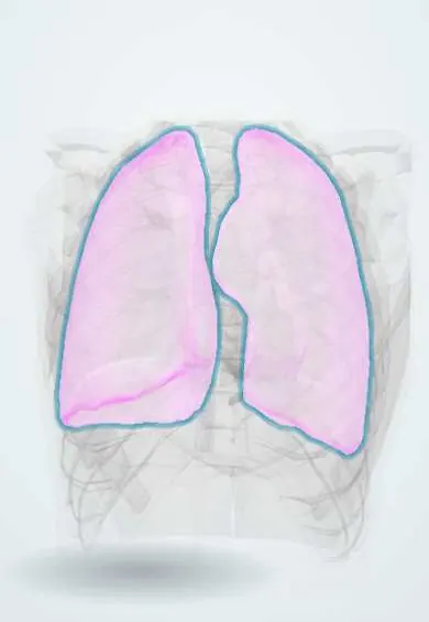
Representative model, for educational purposes only.