Pneumoconioses (silicosis, asbestosis, coal workers' pneumoconiosis) !
Pneumoconioses are lung diseases caused by inhaling mineral dust at work, such as silica, coal or asbestos. They scar the lungs and cannot be cured.
Updated 2026-11-01
Structures involved
Tap one to see what it is and open it in 3D.
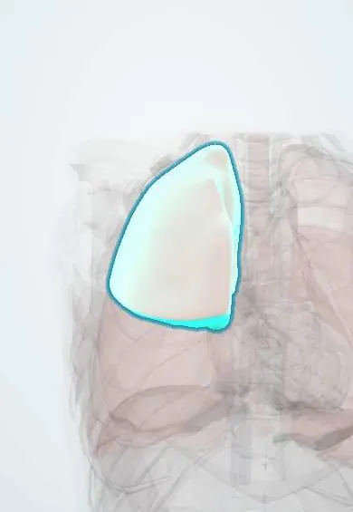
- Right upper lobe
- Left upper lobe
- Right lower lobe
- Pleura
- Left lower lobe
- Mediastinal lymph nodes
- Diaphragm
- Right ventricle
- Pulmonary arteries
Why it happens
- Fibrosis in organs: TGF-β and the myofibroblast
- Air pollution and occupational exposures
- Pigments in tissue: lipofuscin, hemosiderin, melanin, bilirubin, carbon
- Chemical carcinogens: initiation, promotion and the table to know
- Chronic inflammation: causes, cells and features
- Obstructive and restrictive patterns, and flow–volume loops
What it is
A group of occupational interstitial lung diseases caused by inhaling and retaining inorganic dust, which triggers inflammation and fibrosis. The main ones are silicosis (crystalline silica), coal workers' pneumoconiosis (coal dust) and asbestosis (asbestos fibres).
Who gets it
A priority occupational disease in Colombia, where coal mining (Boyacá, Cundinamarca, Norte de Santander, Cesar, La Guajira), sandblasting, stone cutting, brick making, foundries and construction expose many workers to dust, often in small or informal operations. Asbestos was widely used in roofing sheets and pipes and its use was banned in Colombia by law in 2019; disease from past exposure still appears decades later.
Causes
- Silicosis: crystalline silica in mining, quarrying, sandblasting, stone and glass work, ceramics and construction
- Coal workers' pneumoconiosis (black lung): coal mine dust
- Asbestosis: asbestos fibres in mining, insulation, brake linings, cement sheets, shipyards and demolition
- Others: berylliosis (granulomatous, aerospace and electronics), siderosis (iron), talcosis
Risk factors
- Years of exposure and dust concentration
- Work without ventilation, wet methods or respiratory protection
- Smoking (multiplies the risk of lung cancer with asbestos)
- Informal or small-scale mining
Pathophysiology
Small inhaled particles reach the alveoli and are engulfed by macrophages. Silica is toxic to macrophages, which die and release cytokines and growth factors that drive fibroblasts, forming fibrotic nodules; damaged macrophages also impair defence against mycobacteria. Coal dust forms macules and nodules around respiratory bronchioles. Asbestos fibres are too long to be cleared, causing chronic inflammation and diffuse fibrosis. Silica and coal predominate in the upper lobes; asbestosis predominates in the lower lobes and pleura.
Classification
- Silicosis: chronic simple (after 10 or more years), accelerated (5–10 years), acute silicoproteinosis (weeks to few years after massive exposure)
- Simple versus complicated (progressive massive fibrosis, conglomerate masses over 1 cm)
- Upper-lobe pattern: silicosis, coal; lower-lobe pattern: asbestosis
- ILO classification standardises reading of chest X-rays
Symptoms and signs
- No symptoms in early disease, found on screening X-ray
- Progressive exertional shortness of breath
- Chronic cough, with black sputum in coal miners
- Fine basal "velcro" crackles and digital clubbing in asbestosis
- Signs of right heart failure in advanced disease
Diagnosis
- Clinical
- Based on a detailed occupational history (job, years, dust, protection) together with compatible imaging and exclusion of other causes; biopsy is rarely needed. Occupational diagnosis has legal and compensation implications in Colombia.
- Tests
- Spirometry and DLCO (restrictive pattern or mixed; low diffusing capacity); oximetry and walk test. Tuberculosis screening (tuberculin test or interferon-gamma release assay, sputum testing when symptomatic) in silica-exposed workers. Autoantibodies when rheumatoid nodules are suspected (Caplan syndrome).
- Imaging
- Chest X-ray read with the ILO classification: small upper-lobe nodules and eggshell calcification of hilar nodes in silicosis; lower-lobe reticulation and calcified pleural plaques in asbestosis. High-resolution CT is more sensitive and shows conglomerate masses.
Differential diagnosis
- Pulmonary tuberculosis
- Sarcoidosis
- Idiopathic pulmonary fibrosis and other interstitial lung diseases
- Hypersensitivity pneumonitis
- Lung cancer or metastases
Goal of treatment
Stop further exposure, slow progression, detect and treat tuberculosis and cancer early, and maintain function and quality of life; no treatment reverses established fibrosis.
Treatment
- Lifestyle
- Removal from exposure, change of job when possible, and stopping smoking.
- Medical
- Treatment of airflow limitation with bronchodilators, vaccination against influenza and pneumococcus, and treatment of latent or active tuberculosis.
- Rehabilitation
- Pulmonary rehabilitation and long-term oxygen therapy when chronic hypoxaemia is present.
- Procedure
- Whole-lung lavage in acute silicoproteinosis in specialised centres; lung transplantation in selected end-stage patients.
Complications
- Tuberculosis and non-tuberculous mycobacterial infection (especially silicosis)
- Lung cancer (silica and asbestos) and mesothelioma (asbestos)
- Progressive massive fibrosis and respiratory failure
- Pulmonary hypertension and cor pulmonale
- Autoimmune disease associated with silica (rheumatoid arthritis, systemic sclerosis)
Prognosis
Simple disease often remains stable for years after exposure stops. Complicated disease (progressive massive fibrosis), acute silicosis and asbestosis can progress even without further exposure and shorten life, mainly through respiratory failure, tuberculosis and cancer.
Prevention
- Dust control at the source: wet cutting, ventilation, enclosure and substitution of materials
- Certified respiratory protection and training
- Periodic medical surveillance with spirometry and ILO-classified chest X-rays
- Ban on asbestos and safe removal of existing materials
Test yourself
Think of the answer, then tap the question to see it.
Which pneumoconioses predominate in the upper lobes and which in the lower lobes?
Silicosis and coal workers' pneumoconiosis affect mainly the upper lobes; asbestosis affects mainly the lower lobes and pleura.
Which infection is classically increased in patients with silicosis?
Tuberculosis, because silica damages macrophages and impairs defence against mycobacteria.
What chest imaging finding suggests past asbestos exposure?
Calcified pleural plaques, especially on the diaphragm and lower chest wall.
What is eggshell calcification and in which disease is it typical?
Peripheral calcification of hilar and mediastinal lymph nodes, typical of silicosis.
What is the single most important measure once a pneumoconiosis is diagnosed?
Ending exposure to the dust, together with stopping smoking.
Related conditions
- Pulmonary tuberculosis
- Lung cancer
- Malignant pleural mesothelioma
- Interstitial lung disease
- Chronic obstructive pulmonary disease (COPD)
- Rheumatoid arthritis
- Sarcoidosis
- Hypersensitivity pneumonitis
- Pulmonary hypertension and cor pulmonale
Educational information, not medical advice. For any symptom, see a health professional.
Sources: Silicosis (MedlinePlus Medical Encyclopedia (U.S. National Library of Medicine), 2024); Coal worker's pneumoconiosis (MedlinePlus Medical Encyclopedia (U.S. National Library of Medicine), 2024); Asbestosis (MedlinePlus Medical Encyclopedia (U.S. National Library of Medicine), 2024).
Spotted a mistake?We are always improving for the community. Tell us what is wrong and our team will review it.Report a mistake
Representative model, for educational purposes only.