Conditions · Hematology and oncology
Superior vena cava syndrome !
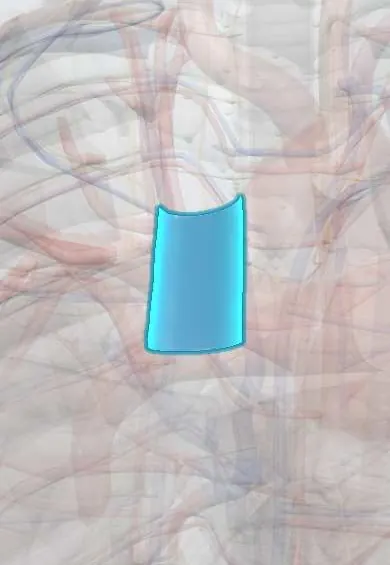
The large vein that drains the head, neck and arms into the heart is squeezed or blocked, usually by a chest tumour, so the face, neck and arms swell.
Updated 2026-11-01
Structures involved
Tap one to see what it is and open it in 3D.
- Superior vena cava
- Mediastinal lymph nodes
- Brachiocephalic veins
- Azygos veins
- Trachea
- Main bronchi
- Right atrium
- Thymus
Why it happens
- Edema and effusions: the four mechanisms
- Local effects of tumours and cancer cachexia
- Starling forces at the capillary
- Central venous pressure and the jugular pulse
What it is
Narrowing or blockage of the superior vena cava (SVC), the thin-walled vein that drains the head, neck and arms into the right atrium, usually by a mediastinal tumour or a clot. It is an oncologic emergency mainly when the airway or brain is affected.
Who gets it
Uncommon overall but a classic oncology presentation: most cases are due to malignancy, above all non-small-cell and small-cell lung cancer, followed by non-Hodgkin lymphoma. Benign causes, chiefly thrombosis around central venous catheters, pacemaker leads and dialysis lines, are increasing. In Colombia and Latin America, where lung cancer is often diagnosed late and tuberculosis and histoplasmosis are present, mediastinal tumours and fibrosing mediastinitis are both causes to consider.
Causes
- Malignant (most cases): lung cancer, especially small-cell and right upper lobe tumours, and mediastinal lymphoma
- Other malignancies: thymoma, germ cell tumours, metastatic lymph nodes (e.g. breast cancer)
- Thrombosis of the SVC from central venous catheters, ports, pacemaker or defibrillator leads
- Fibrosing mediastinitis after histoplasmosis or tuberculosis, or after mediastinal radiotherapy
- Other compression: aortic aneurysm, large retrosternal goitre, constrictive pericarditis
Risk factors
- Smoking and known or suspected lung cancer
- Lymphoma with bulky mediastinal disease
- Long-term central venous catheters, ports or haemodialysis lines
- Pacemaker or implantable defibrillator leads
- Previous chest radiotherapy or granulomatous chest infection
Pathophysiology
External compression, tumour invasion or a clot raises venous pressure in the head, neck and arms. Blood is forced through collateral veins (azygos, internal mammary, chest wall veins), which dilate over days to weeks; slow obstruction therefore causes milder symptoms than sudden thrombosis. Venous congestion produces oedema of the face, larynx and brain, and reduced venous return can lower cardiac output. A mass large enough to block the SVC may also compress the trachea, causing stridor.
Classification
- By cause: malignant or benign (thrombotic, fibrotic, other compression)
- By severity (graded scales such as the Yale system): from asymptomatic on imaging, to mild-moderate oedema, to severe oedema, to life-threatening cerebral oedema, laryngeal oedema or haemodynamic compromise
- By speed: gradual (with collaterals) or acute (often thrombotic)
Symptoms and signs
- Swelling of the face, neck and eyelids, often worse in the morning or when lying down or bending forward
- Breathlessness, the most common symptom, with cough
- Swelling of one or both arms
- Distended neck veins and visible dilated veins over the upper chest
- Reddish or bluish (plethoric) face, headache and a feeling of fullness in the head
- Hoarseness or difficulty swallowing
Diagnosis
- Clinical
- Mainly clinical: facial and arm swelling with distended neck and chest wall veins in a patient with a chest mass, cancer risk or central line. Pemberton's manoeuvre (raising both arms) may worsen facial flushing. Assess airway and level of consciousness first.
- Tests
- A tissue diagnosis is essential before cancer treatment when the patient is stable: sputum cytology, biopsy of an accessible lymph node, bronchoscopy, or CT-guided or mediastinoscopic biopsy. Blood count, LDH and other tests guide the search for lymphoma, germ cell tumour or small-cell cancer. Avoid inserting lines in the arms on the affected side.
- Imaging
- Chest X-ray often shows a widened mediastinum or right hilar mass. Contrast-enhanced CT of the chest is the key test: it shows the level and cause of the obstruction, clot and collaterals. Doppler ultrasound of the arm and neck veins, and venography when stenting is planned.
Differential diagnosis
- Cardiac tamponade or constrictive pericarditis (raised neck veins, but with pulsus paradoxus and no facial oedema pattern)
- Right heart failure
- Angioedema or allergic reaction
- Isolated subclavian or axillary vein thrombosis
- Nephrotic syndrome or other causes of facial oedema
Goal of treatment
Protect the airway and brain, relieve the obstruction quickly when severe, and treat the underlying cause after obtaining a tissue diagnosis.
Treatment
- Acute
- Supportive care: sit the patient upright, give oxygen as needed and secure the airway when there is stridor; corticosteroids and loop diuretics are sometimes used for short-term relief, though their benefit is uncertain except in steroid-responsive tumours such as lymphoma.
- Procedure
- Endovascular stenting of the SVC gives the fastest symptom relief and is the first choice for severe or life-threatening obstruction, often before the cause is fully defined.
- Medical
- Cause-specific cancer treatment: chemotherapy for chemosensitive tumours (small-cell lung cancer, lymphoma, germ cell tumours); radiotherapy for other malignancies.
- Medical
- Catheter-related thrombosis: anticoagulation, with catheter removal or catheter-directed thrombolysis in selected cases.
- Surgical
- Surgical bypass or reconstruction is rarely used, mainly for benign causes such as fibrosing mediastinitis.
- Follow-up
- Nursing priorities: head of bed elevated, watch respiratory rate, stridor, oxygen saturation and level of consciousness, avoid blood pressure cuffs and IV lines in the affected arms, and monitor for bleeding after stenting or on anticoagulants.
Complications
- Laryngeal oedema and airway obstruction
- Cerebral oedema with confusion, seizures or coma
- Haemodynamic compromise
- Extension of thrombosis and pulmonary embolism
- Stent migration, re-occlusion or bleeding
Prognosis
Symptoms usually improve within days after stenting, chemotherapy or radiotherapy. Long-term outlook depends on the underlying disease: it is good for benign causes and lymphoma, but most patients with SVC syndrome from lung cancer have advanced disease and limited survival.
Prevention
- Early diagnosis of lung cancer and lymphoma
- Careful placement, care and timely removal of central venous catheters
- Smoking cessation
Test yourself
Think of the answer, then tap the question to see it.
What is the most common cause of superior vena cava syndrome?
Malignancy, above all lung cancer (especially small-cell), followed by non-Hodgkin lymphoma.
Which imaging test best defines the level and cause of SVC obstruction?
Contrast-enhanced CT of the chest.
Which two findings make SVC syndrome a life-threatening emergency?
Stridor from laryngeal oedema or tracheal compression, and confusion or reduced consciousness from cerebral oedema.
Which treatment gives the fastest relief in severe SVC syndrome?
Endovascular stenting of the superior vena cava.
Why should IV lines be avoided in the arms of a patient with SVC syndrome?
Venous return from the arms is blocked, so infusions drain poorly, worsen oedema and raise the risk of thrombosis.
Related conditions
- Lung cancer
- Non-Hodgkin lymphoma
- Hodgkin lymphoma
- Deep vein thrombosis
- Pulmonary tuberculosis
- Cardiac tamponade
- Heart failure
- Pleural effusion
- Malignant spinal cord compression
- Tumour lysis syndrome
Educational information, not medical advice. For any symptom, see a health professional.
Sources: SVC obstruction (MedlinePlus (U.S. National Library of Medicine), 2025).
Spotted a mistake?We are always improving for the community. Tell us what is wrong and our team will review it.Report a mistake
Representative model, for educational purposes only.