Conditions · Nephrology and urology
Renal vein thrombosis !
A blood clot in the vein that drains a kidney. In adults it is linked to nephrotic syndrome; in newborns to dehydration.
Updated 2026-11-01
Structures involved
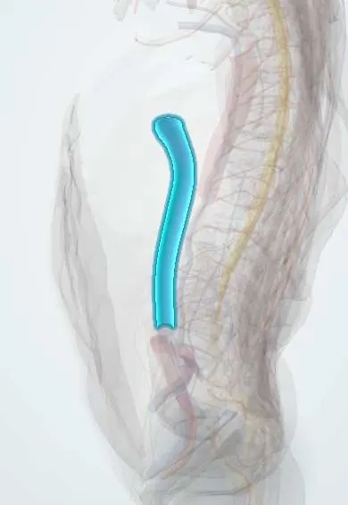
Tap one to see what it is and open it in 3D.
- Left renal vein
- Right renal vein
- Inferior vena cava
- Kidneys
- Testicular vessels
- Left adrenal gland
- Abdominal aorta
Why it happens
- Thrombosis and Virchow's triad
- Hypercoagulable states: inherited and acquired
- Fibrinolysis and natural anticoagulants
What it is
Thrombosis of one or both renal veins, which may extend into the inferior vena cava. It can be acute, with pain, haematuria and loss of function, or chronic and silent, found while evaluating nephrotic syndrome or pulmonary embolism.
Who gets it
Uncommon. In adults most cases occur with nephrotic syndrome, above all membranous nephropathy, where it is found in a substantial minority on imaging. In newborns it is the most common non-catheter-related venous thrombosis, linked to dehydration, perinatal asphyxia, maternal diabetes and sepsis.
Causes
- Nephrotic syndrome, especially membranous nephropathy (loss of antithrombin and other anticoagulant proteins)
- Renal cell carcinoma growing into the renal vein
- Severe dehydration in infants (for example from diarrhoea)
- Trauma, compression of the vein or kidney transplantation
- Inherited or acquired thrombophilia, antiphospholipid syndrome, oral contraceptives and pregnancy
Risk factors
- Nephrotic-range proteinuria with low serum albumin
- Membranous nephropathy
- Dehydration and sepsis in newborns
- Thrombophilia or previous venous thromboembolism
- Kidney tumour invading the vein
Pathophysiology
The process follows Virchow triad. In nephrotic syndrome the kidney leaks antithrombin III and other small anticoagulant proteins while the liver makes more fibrinogen and clotting factors; added haemoconcentration from diuretics makes the blood hypercoagulable. A clot in the renal vein raises back-pressure in the kidney, causing congestion, swelling, haemorrhage and reduced filtration. The left renal vein is longer and receives the left gonadal vein, so left-sided thrombosis can cause a left varicocele in men. Fragments can embolise to the lungs.
Classification
- Acute (pain, haematuria, loss of function) or chronic (often silent, collateral veins develop)
- Unilateral or bilateral; with or without extension into the inferior vena cava
- Neonatal or adult
Symptoms and signs
- Sudden flank or lower back pain
- Gross or microscopic haematuria
- Reduced urine output and worsening kidney function
- An enlarged, palpable kidney in a newborn, with haematuria and low platelets
- Sudden shortness of breath or chest pain if a pulmonary embolism occurs
- Often no symptoms in chronic cases
Diagnosis
- Clinical
- Suspect it with sudden flank pain and haematuria, worsening proteinuria or kidney function in nephrotic syndrome, a new left varicocele or pulmonary embolism in a nephrotic patient.
- Tests
- Urinalysis (haematuria, proteinuria), creatinine, serum albumin and urine protein; D-dimer is supportive but non-specific. Thrombophilia testing in selected patients.
- Imaging
- Contrast CT or MR venography is the usual confirmation; Doppler ultrasound is used first in newborns and shows an enlarged kidney with absent venous flow. Catheter venography is the reference standard but rarely needed.
Differential diagnosis
- Kidney stones
- Acute pyelonephritis
- Renal infarction from arterial embolism
- Renal cell carcinoma
- Worsening of the underlying glomerular disease
Goal of treatment
Stop the clot from growing or embolising, protect kidney function and treat the underlying cause.
Treatment
- Medical
- Anticoagulation, initially with heparins and then with oral anticoagulants, usually for months and for as long as the nephrotic syndrome persists.
- Medical
- Treatment of the cause: immunosuppressive therapy for membranous nephropathy, renin–angiotensin blockers to reduce proteinuria; prophylactic anticoagulation is considered in high-risk nephrotic patients.
- Procedure
- Catheter-directed thrombolysis or thrombectomy in selected patients with acute bilateral thrombosis, acute kidney injury or extension into the vena cava; nephrectomy with thrombectomy for tumour thrombus.
- Acute
- In newborns: correction of dehydration and electrolytes, treatment of sepsis and supportive care, with anticoagulation in selected cases.
- Follow-up
- Monitoring of kidney function, platelet count and bleeding during anticoagulation.
Complications
- Pulmonary embolism
- Acute kidney injury
- Chronic kidney disease or atrophy of the affected kidney
- Extension into the inferior vena cava
- Hypertension (especially after neonatal thrombosis)
- Bleeding from anticoagulation
Prognosis
Most adults improve with anticoagulation and treatment of the underlying disease, and lasting kidney damage is uncommon. Death is more often due to pulmonary embolism or the underlying condition. Neonatal cases can leave an atrophic kidney and hypertension.
Prevention
- Preventing dehydration in infants with diarrhoea
- Assessing clotting risk and considering prophylaxis in severe nephrotic syndrome
Test yourself
Think of the answer, then tap the question to see it.
Which glomerular disease is most associated with renal vein thrombosis?
Membranous nephropathy.
Why is nephrotic syndrome hypercoagulable?
Urinary loss of antithrombin III and other anticoagulant proteins plus increased hepatic synthesis of fibrinogen and clotting factors.
Why can left renal vein thrombosis cause a varicocele?
The left gonadal vein drains into the left renal vein, so obstruction raises pressure in the pampiniform plexus.
Classic neonatal presentation?
A flank mass, haematuria and thrombocytopenia in a dehydrated or septic newborn.
Related conditions
- Nephrotic syndrome
- Membranous nephropathy
- Pulmonary embolism
- Deep vein thrombosis
- Inherited thrombophilia
- Antiphospholipid syndrome
- Renal cell carcinoma
- Acute kidney injury
- Varicocele
- Kidney transplant rejection
- Acute (viral) gastroenteritis
Educational information, not medical advice. For any symptom, see a health professional.
Sources: Renal vein thrombosis: MedlinePlus Medical Encyclopedia (MedlinePlus (U.S. National Library of Medicine), 2024); Nephrotic syndrome: MedlinePlus Medical Encyclopedia (MedlinePlus (U.S. National Library of Medicine), 2024).
Spotted a mistake?We are always improving for the community. Tell us what is wrong and our team will review it.Report a mistake
Representative model, for educational purposes only.