Cardiovascular system · Abdomen
Testicular vessels !
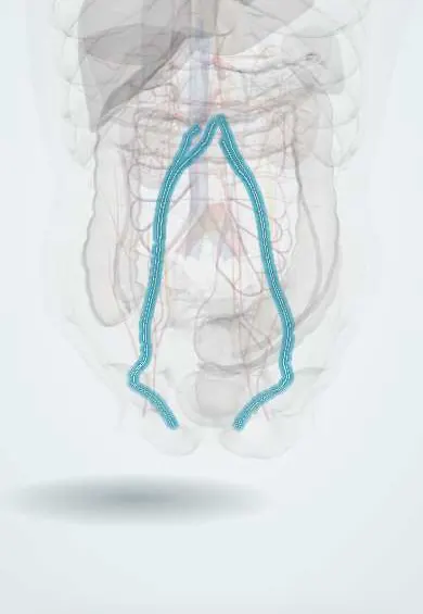
The gonadal vessels are the long arteries and veins that run down the back of the abdomen to the testes or ovaries; testicular lymph follows them upward.
Official name: Testicular artery
Opens in the Durgical app, no sign-up needed.
What it is
Long thin arteries and veins running down the back wall of the abdomen to the testes (male base model; the ovarian vessels in women).
What it does
Supply the testes. The left vein drains into the left renal vein, which is why varicoceles are usually on the left.
Key facts
- Origin
- Testicular (ovarian) arteries: anterior aorta at L2, below renal arteries; run down on psoas and cross the ureter
- Branches
- Ureteric branches; enter spermatic cord at deep inguinal ring (ovarian: suspensory ligament of ovary)
- Supplies
- Testis and epididymis (ovary, uterine tube); veins: pampiniform plexus, right into IVC, left into left renal vein
Relations
- Course
- From the aorta at L2 they run down on the psoas major behind the peritoneum, cross in front of the ureter and the external iliac artery and pass through the deep inguinal ring into the spermatic cord (ovarian vessels: over the pelvic brim into the suspensory ligament of the ovary)
- In front
- Right: third part of the duodenum, ileocolic and right colic vessels and the root of the mesentery; left: left colic vessels and the sigmoid colon
- Behind
- Psoas major, genitofemoral nerve, the ureter (crossed obliquely) and lower down the external iliac vessels
How to find it
Seen once the posterior peritoneum is opened: they run down on the front of psoas major and cross in front of the ureter; the testicular vessels then reach the deep inguinal ring, while the ovarian vessels cross the pelvic brim inside the suspensory ligament of the ovary. In the scrotum the pampiniform plexus is felt as part of the spermatic cord.
Examination
- Varicocele examination, standing and then with a Valsalva manoeuvre: a soft mass like a 'bag of worms' above the testis, or one that appears only on straining, means dilated pampiniform veins; it should shrink when the person lies down
On imaging
Scrotal Doppler ultrasound shows a varicocele as dilated veins of the pampiniform plexus with reversed flow on Valsalva. On CT the gonadal veins run down beside the ureter; ovarian vein thrombosis appears as a tubular filling defect, more often on the right after childbirth.
Clinical notes
- A new right-sided varicocele, or one that does not empty lying down, raises suspicion of a mass pressing on the inferior vena cava or renal vein
- Compression of the left renal vein between the aorta and the superior mesenteric artery (nutcracker) can cause a left varicocele
- In oophorectomy the ureter can be caught when the suspensory ligament with the ovarian vessels is clamped at the pelvic brim
- Reflux in the ovarian veins is linked to pelvic congestion syndrome
- Short testicular vessels can limit bringing down an undescended testis; one staged technique divides them and relies on the artery of the ductus deferens
Anatomical variants
- The testicular artery may arise from the renal artery or an accessory renal artery instead of the aorta
- The right testicular vein occasionally drains into the right renal vein
Development
The gonads form on the posterior abdominal wall near the upper lumbar levels and descend, taking their vessels with them: this explains the long course, the origin at L2 and the para-aortic lymph drainage.
Test yourself
Think of the answer, then tap the question to see it.
Where do the testicular arteries arise and run?
From the front of the aorta at L2, below the renal arteries; they run down on psoas major, cross in front of the ureter and enter the spermatic cord at the deep inguinal ring.
Why are varicoceles usually on the left?
The left testicular vein is longer and joins the left renal vein at a right angle, while the right enters the inferior vena cava obliquely.
To which lymph nodes does the testis drain first, and why?
To the para-aortic nodes near the renal veins, not the inguinal nodes, because the testis formed on the posterior abdominal wall and descended with its vessels.
What does a new right-sided varicocele, or one that does not empty lying down, raise suspicion of?
A mass pressing on the inferior vena cava or renal vein.
Educational information, not medical advice. For any symptom, see a health professional.
Its names
- Official Latin (TA2)
- arteria testicularis; vena testicularis sinistra; vena testicularis dextra TA2 4288
- Official Spanish (DeCS)
- Vasos testiculares
- English (TA2)
- Testicular artery
- Also called
- internal spermatic artery · arteria spermatica interna · arteria spermatica
Related
Sources: Terminologia Anatomica (TA2); Anatomy, Abdomen and Pelvis: Arteries; Anatomy of the Human Body (20th ed.): The Abdominal Aorta (Lea & Febiger (Gray's Anatomy, 1918), 1918) · adapted, Public domain.
Spotted a mistake?We are always improving for the community. Tell us what is wrong and our team will review it.Report a mistake
Representative model, for educational purposes only.