Conditions · Nephrology and urology
Rhabdomyolysis !
Rhabdomyolysis is the rapid breakdown of skeletal muscle. Its contents leak into the blood and can damage the kidneys and upset potassium levels.
Updated 2026-11-01
Structures involved
Tap one to see what it is and open it in 3D.
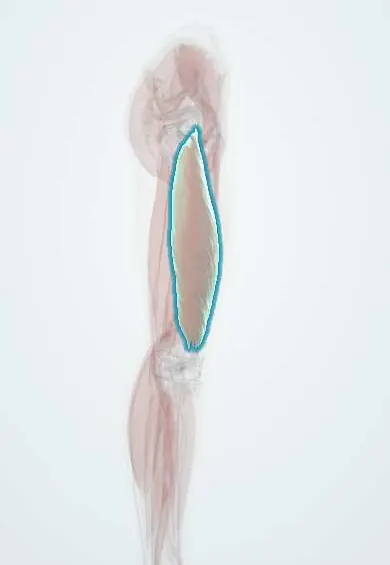
- Vastus lateralis
- Gastrocnemius (inner head)
- Kidneys
- Rectus femoris
- Vastus medialis
- Gastrocnemius (outer head)
- Soleus
- Gluteus maximus
- Vessels inside the kidney
In biochemistry
In the clinical exam
Why it happens
- Mechanisms of cell injury: ATP, mitochondria, calcium, membranes and DNA
- Thermal injury: burns, hyperthermia and hypothermia
- Mechanical and electrical injury: abrasion, contusion, laceration, wounds
- Reversible vs irreversible cell injury: the point of no return
- Drugs of abuse: cocaine, opioids, amphetamines, cannabis
- Physiology of acute and chronic kidney failure
Show 4 more
What it is
A syndrome in which injured skeletal muscle releases its contents (myoglobin, potassium, phosphate, creatine kinase) into the circulation. Its main danger is acute kidney injury and life-threatening hyperkalaemia.
Who gets it
Seen in trauma and crush injuries, after extreme exertion (military training, marathons), in heat illness and with drugs and toxins. In Colombia and Latin America, snakebite envenomation (especially some rattlesnake and coral-snake species), earthquakes and building collapse, and traffic trauma are relevant causes.
Causes
- Trauma and crush injury, prolonged immobilisation (a person found on the floor after a fall, coma, long surgery)
- Severe or unaccustomed exertion, especially in heat or with dehydration
- Heat stroke, malignant hyperthermia and neuroleptic malignant syndrome
- Drugs and toxins: alcohol, cocaine and amphetamines, statins (rarely, more with interacting drugs), some snake venoms
- Infections (influenza, other viruses, sepsis) and electrolyte disorders such as severe hypokalaemia or hypophosphataemia
- Inherited muscle disorders (metabolic myopathies) and prolonged seizures
Risk factors
- Intense exercise without conditioning, heat and poor hydration
- Alcohol or stimulant use
- Statin therapy combined with interacting drugs
- Older age and immobility after a fall
- Inherited myopathy or previous episodes
Pathophysiology
Injury or energy failure in the muscle cell lets calcium flood in, activating enzymes that destroy the membrane. Myoglobin, potassium, phosphate and urate pour into the blood. In the kidney, myoglobin is toxic to tubular cells, forms casts that block the tubules (worse in acidic urine) and causes vasoconstriction, while fluid trapped in injured muscle drops the circulating volume.
Classification
- Traumatic (crush, compartment syndrome) or non-traumatic (exertional, toxic, metabolic, infectious, inherited)
- Uncomplicated or complicated by acute kidney injury, hyperkalaemia or compartment syndrome
Symptoms and signs
- Muscle pain, tenderness and swelling, most often in the thighs, calves or lower back
- Muscle weakness
- Dark, tea- or cola-coloured urine
- Reduced urine output
- Fatigue, nausea, fever or confusion depending on the cause
- The classic triad (pain, weakness, dark urine) appears in a minority of patients
Diagnosis
- Clinical
- History of trauma, immobilisation, exertion, heat, drugs or envenomation, with muscle pain and dark urine. Check limbs for compartment syndrome and monitor urine output.
- Tests
- Creatine kinase (CK) is the key test: markedly raised, commonly taken as more than five times the upper limit, peaking within a few days. Urine dipstick positive for blood with few or no red cells on microscopy (myoglobin). Potassium, calcium, phosphate, creatinine, urea, uric acid, blood gas, coagulation, and an ECG for hyperkalaemia.
- Imaging
- Not needed to diagnose. Imaging or compartment pressure measurement when compartment syndrome is suspected; imaging for associated trauma.
Differential diagnosis
- Haematuria from urinary tract disease (red cells present on microscopy)
- Haemolysis with haemoglobinuria
- Inflammatory myositis (polymyositis, dermatomyositis)
- Benign exercise-related CK rise without kidney involvement
- Other causes of acute kidney injury
Goal of treatment
Restore circulating volume early to protect the kidneys, correct dangerous electrolyte disturbances, remove the cause and detect complications such as compartment syndrome.
Treatment
- Acute
- Early and generous intravenous isotonic fluids, aiming for a high urine output; this is the cornerstone of treatment.
- Acute
- Urgent treatment of hyperkalaemia (cardiac membrane stabilisation with calcium, shifting potassium into cells, removal); low calcium is usually not corrected unless symptomatic or with hyperkalaemia, because it can rebound.
- Follow-up
- Nursing priorities: strict intake and output (often with a urinary catheter), urine colour, cardiac monitoring, serial CK and electrolytes, limb checks for swelling and pain.
- Procedure
- Dialysis (kidney replacement therapy) for refractory hyperkalaemia, acidosis, fluid overload or severe kidney failure.
- Surgical
- Fasciotomy for established compartment syndrome; stop offending drugs, cool heat stroke and give antivenom in envenomation.
Complications
- Acute kidney injury (pigment nephropathy)
- Hyperkalaemia with arrhythmias and cardiac arrest
- Early hypocalcaemia and later hypercalcaemia during recovery
- Hyperphosphataemia, hyperuricaemia and metabolic acidosis
- Compartment syndrome
- Disseminated intravascular coagulation
Prognosis
Usually good when treated early; most people recover full kidney function. Outcome is worse with severe acute kidney injury, older age, sepsis or multiple trauma; recurrent episodes suggest an underlying myopathy.
Prevention
- Gradual conditioning, rest and hydration during intense exercise and in hot weather
- Early fluid resuscitation in crush victims, ideally before release from entrapment
- Review of drug interactions in people taking statins
Test yourself
Think of the answer, then tap the question to see it.
Which laboratory test is the key marker of rhabdomyolysis?
Serum creatine kinase (CK), markedly raised (commonly more than five times the upper limit of normal).
Why is the urine dipstick positive for blood with few red cells on microscopy?
The dipstick detects the haem in myoglobin, which is filtered into the urine, while there are no red cells.
What is the cornerstone of initial treatment?
Early, aggressive intravenous isotonic fluids to maintain a high urine output and prevent acute kidney injury.
Which electrolyte disturbance is the most immediately life-threatening?
Hyperkalaemia, released from damaged muscle, which can cause arrhythmias and cardiac arrest.
Related conditions
- Acute kidney injury
- Acute compartment syndrome
- Hyperkalaemia
- Hypocalcaemia
- Heat stroke and heat exhaustion
- Malignant hyperthermia
- Neuroleptic malignant syndrome
- Cocaine and sympathomimetic toxicity
- Snakebite envenomation (Bothrops, Lachesis, Crotalus)
Educational information, not medical advice. For any symptom, see a health professional.
Sources: Rhabdomyolysis: MedlinePlus Medical Encyclopedia (MedlinePlus (U.S. National Library of Medicine), 2024).
Spotted a mistake?We are always improving for the community. Tell us what is wrong and our team will review it.Report a mistake
Representative model, for educational purposes only.