Conditions · Pediatrics and neonatology
Sturge–Weber syndrome !
A birth condition with a facial port-wine stain, abnormal blood vessels over the brain and often glaucoma, causing seizures and weakness.
Updated 2026-11-01
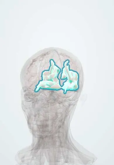
Structures involved
Tap one to see what it is and open it in 3D.
Why it happens
What it is
A sporadic neurocutaneous disorder (phakomatosis) in which capillary-venous malformations affect the facial skin in the territory of the ophthalmic branch of the trigeminal nerve, the leptomeninges over the brain (usually occipital and parietal) and the eye, leading to seizures, stroke-like episodes, developmental problems and glaucoma.
Who gets it
Rare: roughly 1 in 20,000 to 50,000 births, with no sex or ethnic predilection. Not inherited in families. Only a minority (often quoted as about 10 to 30%) of infants with a port-wine stain in the forehead or upper eyelid area turn out to have brain involvement.
Causes
- Somatic (post-zygotic, mosaic) activating variant of the GNAQ gene in developing vascular cells
- Not inherited and not caused by anything during pregnancy; occurs by chance early in embryonic development
- Persistence of a primitive embryonic venous plexus around the cephalic neural tube
Risk factors
- Port-wine stain involving the forehead or upper eyelid (V1 territory) is the main marker of risk
- Bilateral or extensive facial stains carry a higher risk of brain and eye involvement
- No known parental or pregnancy risk factors
Pathophysiology
The GNAQ variant increases signalling in endothelial cells and disturbs vessel maturation. A leptomeningeal angioma covers the cortex, usually on the same side as the facial stain. Venous drainage of the underlying cortex is poor, causing chronic venous congestion, ischaemia, calcification (tram-track pattern) and progressive cortical atrophy. This damaged cortex is epileptogenic and seizures worsen the ischaemia. In the eye, choroidal haemangioma and raised episcleral venous pressure or angle anomalies cause glaucoma.
Classification
- Roach type I: facial and leptomeningeal angioma, with or without glaucoma (classic form)
- Roach type II: facial angioma, with or without glaucoma, without brain involvement
- Roach type III: isolated leptomeningeal angioma, usually without glaucoma
Symptoms and signs
- Port-wine stain (capillary malformation) present at birth, usually on the forehead and upper eyelid, often one-sided; it darkens and may thicken with age
- Focal seizures, often starting in the first year of life, frequently with fever
- Hemiparesis on the side opposite the brain lesion and visual field defect (hemianopia)
- Stroke-like episodes with transient weakness, and headaches resembling migraine
- Developmental delay, intellectual disability, attention and behaviour problems
- Glaucoma on the side of the stain (buphthalmos in infants), sometimes congenital
Diagnosis
- Clinical
- Suspected in any newborn with a port-wine stain involving the forehead or upper eyelid; look for seizures, neurological deficits and eye signs. Early eye examination with intraocular pressure measurement is needed.
- Tests
- Electroencephalogram (EEG) for seizures or slowing over the affected hemisphere; ophthalmological examination with tonometry and fundoscopy, repeated over time; genetic testing of affected tissue for GNAQ is possible but not required.
- Imaging
- Contrast-enhanced brain MRI is the key test, showing leptomeningeal enhancement, cortical atrophy and an enlarged choroid plexus; it may be normal in the first months and need repeating. CT shows gyriform 'tram-track' calcifications in older children.
Differential diagnosis
- Isolated port-wine stain without brain involvement
- Infantile haemangioma (appears after birth, grows and then involutes)
- PHACE syndrome
- Klippel–Trénaunay syndrome and other overgrowth vascular syndromes
- Other neurocutaneous disorders such as neurofibromatosis or tuberous sclerosis
Goal of treatment
Control seizures early, protect the developing brain and vision, and reduce the cosmetic and psychosocial burden of the facial stain.
Treatment
- Medical
- Antiseizure medications for seizures; low-dose aspirin is often used to reduce stroke-like episodes and seizure frequency in specialised centres.
- Procedure
- Pulsed dye laser treatment of the port-wine stain, ideally started in infancy.
- Medical
- Glaucoma care: topical pressure-lowering eye drops (such as beta blockers or prostaglandin analogues) as first step.
- Surgical
- Surgery for drug-resistant glaucoma (goniotomy, trabeculectomy) and for refractory epilepsy (focal resection or hemispherectomy).
- Rehabilitation
- Early rehabilitation: physiotherapy, occupational and speech therapy, and educational support.
- Follow-up
- Lifelong follow-up: regular eye pressure checks, neurological and developmental review; teach families seizure first aid.
Complications
- Drug-resistant epilepsy
- Permanent hemiparesis and visual field loss
- Intellectual disability and behavioural or learning problems
- Vision loss from glaucoma or choroidal haemangioma with retinal detachment
- Soft-tissue and bony overgrowth of the face; bleeding from nodular stains
- Headaches and depression or anxiety
Prognosis
Very variable. Children whose seizures start late and are well controlled may have normal intelligence; early-onset, frequent or bilateral disease predicts more neurological impairment. Life expectancy is usually near normal with good seizure and eye care.
Test yourself
Think of the answer, then tap the question to see it.
What is the classic triad of Sturge–Weber syndrome?
Facial port-wine stain in the V1 distribution, ipsilateral leptomeningeal angioma, and glaucoma.
What gene is involved and is it inherited?
A somatic mosaic activating variant of GNAQ; it occurs sporadically and is not inherited.
What is the typical CT finding in an older child?
Gyriform 'tram-track' cortical calcifications, with atrophy of the affected hemisphere.
Which eye complication must be screened for in an infant with a forehead port-wine stain?
Glaucoma (raised intraocular pressure, possibly buphthalmos), on the same side as the stain.
What is the most common neurological presentation?
Focal seizures beginning in infancy, sometimes followed by hemiparesis on the opposite side.
Related conditions
- Epilepsy and seizures
- Open-angle glaucoma
- Infantile hemangioma
- Neurofibromatosis (types 1 and 2)
- Tuberous sclerosis complex
- Migraine
- Ischaemic stroke
Educational information, not medical advice. For any symptom, see a health professional.
Sources: Sturge-Weber syndrome (MedlinePlus Genetics (U.S. National Library of Medicine), 2024).
Spotted a mistake?We are always improving for the community. Tell us what is wrong and our team will review it.Report a mistake
Representative model, for educational purposes only.