Conditions · Pediatrics and neonatology
X-linked (Bruton) agammaglobulinaemia !
An inherited immune disorder in boys who make almost no B cells or antibodies, so they get repeated bacterial infections after maternal antibodies fade.
Updated 2026-11-01
Structures involved
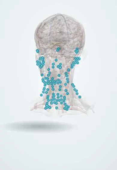
Tap one to see what it is and open it in 3D.
Why it happens
- Mendelian inheritance: autosomal dominant, recessive and X-linked
- Innate and adaptive immunity: overview
- Immunoglobulins (antibodies)
What it is
A primary humoral immunodeficiency caused by mutations in Bruton tyrosine kinase (BTK), which arrests B-cell development at the pre-B stage. Affected boys have almost no circulating B cells and very low levels of all immunoglobulin classes.
Who gets it
Rare: about 1 in 200,000 live births, almost exclusively in males. Symptoms usually start between 6 and 12 months of age. In Latin America, registries of the Latin American Society for Immunodeficiencies show that many cases are diagnosed years after the first infections.
Causes
- Mutations in the BTK gene on the X chromosome
- X-linked recessive: carrier mothers pass it to half of their sons
- About half of patients have no family history (new mutation)
Risk factors
- Male sex
- Brothers or maternal male relatives with recurrent infections or early deaths from infection
- Mother known to be a carrier
Pathophysiology
Without BTK, pre-B cells in the bone marrow cannot mature, so there are no mature B cells, no plasma cells and no antibodies. T-cell immunity is intact. Newborns are protected by maternal IgG that crossed the placenta; once it wanes after about six months, infections by encapsulated bacteria (pneumococcus, Haemophilus influenzae), enteroviruses and Giardia appear. Lymphoid tissues that depend on B cells (tonsils, lymph-node follicles) are tiny or absent.
Symptoms and signs
- Recurrent bacterial otitis media, sinusitis and pneumonia from about 6 months of age
- Infections by encapsulated bacteria (Streptococcus pneumoniae, Haemophilus influenzae)
- Absent or very small tonsils and lymph nodes despite repeated infections
- Chronic diarrhoea and malabsorption, often from Giardia
- Failure to thrive
- Severe or chronic enteroviral infection (meningoencephalitis), and vaccine-associated polio with oral polio vaccine
Diagnosis
- Clinical
- Suspected in a boy with recurrent bacterial infections starting after the first months of life, a family history of affected maternal male relatives, and scant lymphoid tissue on examination.
- Tests
- Serum immunoglobulins show very low IgG, IgA and IgM (interpret with age-specific ranges); absent vaccine antibody responses. Flow cytometry shows CD19+ B cells below 2% with normal T cells. Confirmation by absent BTK protein or BTK gene sequencing; carrier testing of the mother.
- Imaging
- Chest imaging or CT for pneumonia and bronchiectasis; sinus imaging when indicated. A lateral neck radiograph may show absent adenoid tissue.
Differential diagnosis
- Transient hypogammaglobulinaemia of infancy (B cells present, recovers spontaneously)
- Common variable immunodeficiency (later onset, B cells usually present)
- Severe combined immunodeficiency (T-cell defect, opportunistic infections, earlier onset)
- Secondary antibody loss (nephrotic syndrome, protein-losing enteropathy, malnutrition)
Goal of treatment
Replace the missing antibodies for life, prevent and treat infections early, and protect the lungs from bronchiectasis.
Treatment
- Medical
- Lifelong immunoglobulin replacement, intravenous or subcutaneous, adjusted to trough IgG levels and infections.
- Medical
- Prompt antibiotics for each bacterial infection; prophylactic antibiotics in some patients with recurrent infections or bronchiectasis.
- Follow-up
- Regular lung function and chest imaging, IgG trough levels, growth and infection diary; avoid live vaccines (oral polio, among others) in the patient.
- Lifestyle
- Chest physiotherapy when bronchiectasis is present, hand hygiene, and genetic counselling for the family.
Complications
- Bronchiectasis and chronic lung disease
- Chronic enteroviral meningoencephalitis
- Vaccine-associated paralytic polio
- Sepsis and bacterial meningitis
- Chronic diarrhoea and malnutrition
Prognosis
With early diagnosis and regular immunoglobulin replacement, most patients reach adulthood with good quality of life. Outcome depends on preventing lung damage before diagnosis; chronic enteroviral infection carries a poor prognosis.
Prevention
- Carrier testing and genetic counselling for mothers and female relatives
- Testing of at-risk newborn boys before live vaccines are given
Test yourself
Think of the answer, then tap the question to see it.
Which gene is mutated in Bruton agammaglobulinaemia, and at which stage does B-cell development stop?
BTK (Bruton tyrosine kinase); development arrests at the pre-B-cell stage in the bone marrow.
Why do infections usually start after about 6 months of age?
Because maternal IgG transferred across the placenta protects the infant until it wanes.
Which physical finding suggests XLA in a boy with recurrent infections?
Absent or very small tonsils and lymph nodes.
Which laboratory pattern confirms the diagnosis?
Very low IgG, IgA and IgM with almost absent CD19+ B cells and normal T cells; confirmed by BTK testing.
Which vaccine must be avoided in XLA?
Live vaccines, especially oral polio vaccine, which can cause vaccine-associated paralytic polio.
Related conditions
- Acute otitis media
- Acute rhinosinusitis
- Community-acquired pneumonia
- Bronchiectasis
- Giardiasis
- Bacterial meningitis
- Adenosine deaminase deficiency (ADA-SCID)
- DiGeorge syndrome (22q11.2 deletion syndrome)
- Wiskott–Aldrich syndrome
- Chronic granulomatous disease
Educational information, not medical advice. For any symptom, see a health professional.
Sources: X-linked agammaglobulinemia: MedlinePlus Genetics (MedlinePlus, US National Library of Medicine, 2026); About Primary Immunodeficiency (PI) (Centers for Disease Control and Prevention (CDC), 2026).
Spotted a mistake?We are always improving for the community. Tell us what is wrong and our team will review it.Report a mistake
Representative model, for educational purposes only.