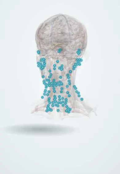
Lymphatic system · Head and neck
Neck lymph nodes !
The neck lymph nodes filter lymph from the head and neck; they swell with infections and are checked closely in head and neck cancer.
Official name: Lymph nodes of neck
Opens in the Durgical app, no sign-up needed.
What it is
Chains of small glands along the jaw, the jugular vein and the back of the neck.
What it does
Filter lymph from the head and neck; they swell with infections and are checked in head and neck cancer.
Key facts
- Where
- Levels I–VI: submental and submandibular, upper, middle and lower jugular chain, posterior triangle, central compartment
- Function
- Drain the head and neck to the jugular lymphatic trunk
Relations
- Deep
- Deep cervical nodes follow the internal jugular vein, along the carotid sheath and under the sternocleidomastoid
- Superficial
- Superficial cervical nodes follow the external jugular vein on the sternocleidomastoid
- Behind
- Level V nodes lie along the accessory nerve in the posterior triangle
- Below
- Lymph collects into the jugular trunk, which ends at the junction of the internal jugular and subclavian veins, joining the thoracic duct on the left or the right lymphatic duct
- Above
- A collar of node groups at the junction of head and neck: occipital, mastoid (retroauricular), parotid (preauricular), submandibular and submental; they drain the scalp and face and empty into the deep jugular and accessory chains
How to find it
Examined from behind with the head bent slightly forward, rolling the nodes under the fingertips in order: submental, submandibular, preauricular, postauricular, occipital, along the sternocleidomastoid, the posterior triangle and the supraclavicular fossa.
On imaging
On ultrasound a benign node is oval with a fatty hilum; a round shape, lost hilum, necrosis, calcification or disorganized peripheral blood flow suggests metastasis. CT, MRI or PET-CT stage head and neck cancer.
Common problems
- Swollen nodes with colds and throat infections, usually tender and temporary.
- Hard, painless, growing nodes for more than a few weeks need checking (lymphoma, cancer spread).
How it is diagnosed
Exam, ultrasound and a needle biopsy when suspicious.
Treatment
- Treat the infection; neck dissection or radiotherapy for cancer.
Clinical notes
- Biopsy of a level V node can injure the accessory nerve, leaving shoulder drop and weak shoulder shrug
- Delphian (prelaryngeal) node on the cricothyroid membrane: when enlarged it suggests laryngeal or thyroid cancer
- Tuberculous lymphadenitis (scrofula): matted, painless nodes that may soften and drain, still common where tuberculosis is frequent
- Neck dissection is radical (levels I–V with the sternocleidomastoid, internal jugular vein and accessory nerve), modified radical (sparing one or more of these) or selective (for example levels I–III)
- Central (level VI) nodes, prelaryngeal, pretracheal and paratracheal chains along the recurrent laryngeal nerves, drain the thyroid, larynx, trachea and esophagus; they are cleared in thyroid cancer surgery, where the recurrent nerve is at risk, and enlarged ones can compress it
Anatomical variants
- Submental nodes drain the chin, central lower lip, lower incisors, floor of the mouth and tongue tip, and may send lymph to the submandibular or deep nodes of both sides, so a midline lip or tongue-tip cancer can spread bilaterally
Related conditions
- Cervical lymphadenopathy
- Thyroid cancer (papillary)
- HIV infection and AIDS
- Hodgkin lymphoma
- Non-Hodgkin lymphoma
- Chronic lymphocytic leukaemia
- Merkel cell carcinoma
- Carcinoma of unknown primary
- Castleman disease
- Infectious mononucleosis
- Paracoccidioidomycosis
- Bartonella infections (cat-scratch disease, Carrion disease)
- Nontuberculous mycobacterial infections (MAC, M. abscessus, Buruli ulcer)
- Human African trypanosomiasis (sleeping sickness)
- Head and neck squamous cell carcinoma (oral cavity and oropharynx, HPV)
- Nasopharyngeal carcinoma
- Kawasaki disease
- X-linked (Bruton) agammaglobulinaemia
- Chronic granulomatous disease
- Hyper-IgM syndrome (CD40 ligand deficiency)
Test yourself
Think of the answer, then tap the question to see it.
Which landmarks separate cervical lymph node levels II, III and IV?
The hyoid bone divides level II from III; the cricoid cartilage (the omohyoid in surgery) divides level III from IV.
What is the Virchow (Troisier) node and what can it signal?
An enlarged left supraclavicular node, where the thoracic duct ends; it may be the first sign of an abdominal cancer, classically of the stomach.
How is a persistent lateral neck lump in an adult treated until proven otherwise?
As metastatic squamous cell carcinoma; the primary is sought in the tonsil, tongue base and nasopharynx.
Which node drains the tonsil and tongue base and enlarges in tonsillitis?
The jugulodigastric node, in level II below the posterior belly of the digastric.
Which nerve is at risk in a level V lymph node biopsy, and what deficit follows?
The accessory nerve: shoulder drop and a weak shoulder shrug.
Educational information, not medical advice. For any symptom, see a health professional.
Its names
- Official Latin (TA2)
- nodi lymphoidei colli TA2 5212
- Official Spanish (DeCS)
- Nódulos linfáticos del cuello DeCS D008198
- English (TA2)
- Lymph nodes of neck
- Also called
- neck glands · cervical lymph nodes
Related
Sources: Terminologia Anatomica (TA2); DeCS; Anatomy, Head and Neck, Lymph Nodes; Anatomy, Head and Neck, Supraclavicular Lymph Node; Tratado de anatomía humana (9.ª ed.), tomo II, Libro IV Angiología, sección 5: Linfáticos.
Spotted a mistake?We are always improving for the community. Tell us what is wrong and our team will review it.Report a mistake
Representative model, for educational purposes only.