Conditions · Gastroenterology and hepatology
Zenker diverticulum (pharyngo-oesophageal diverticulum) !
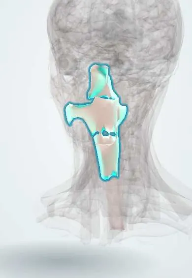
A pouch that bulges out of the back of the throat just above the oesophagus in older adults. Food gets trapped, causing bad breath and regurgitation.
Updated 2026-11-01
Structures involved
Tap one to see what it is and open it in 3D.
Why it happens
- Swallowing and esophageal motility
- The GI tract wall: mucosa, submucosa, muscularis, serosa; enteric plexuses
What it is
An acquired outpouching of the mucosa and submucosa through a weak area in the posterior wall of the lower pharynx, just above the upper oesophageal sphincter (cricopharyngeus). It is a false (pulsion) diverticulum.
Who gets it
Uncommon, seen mainly in adults over 60 and more often in men. It is the most common diverticulum of the upper digestive tract and an important cause of dysphagia and aspiration in older adults.
Causes
- Poor relaxation or reduced compliance of the cricopharyngeus muscle (upper oesophageal sphincter)
- Raised pressure in the lower pharynx during swallowing pushes the mucosa through Killian triangle
- Age-related muscle and connective tissue changes
Risk factors
- Age over 60
- Male sex
- Cricopharyngeal muscle dysfunction or fibrosis
- Possibly gastro-oesophageal reflux
Pathophysiology
When the cricopharyngeus fails to open fully, each swallow raises pressure in the hypopharynx. Over years, the mucosa herniates through Killian triangle, a weak area between the oblique fibres of the inferior pharyngeal constrictor and the horizontal fibres of the cricopharyngeus. The pouch grows, fills with food and saliva, and can compress the oesophagus; the trapped contents later regurgitate into the mouth or are aspirated into the airway.
Classification
- Pulsion (false) diverticulum, unlike traction (true) mid-oesophageal diverticula
- Graded by size on contrast study: small, medium and large pouches
Symptoms and signs
- Oropharyngeal dysphagia that gets worse over time
- Regurgitation of undigested food, sometimes days after eating
- Bad breath (halitosis)
- Gurgling sounds in the neck when swallowing
- Cough, choking or a feeling of a lump in the throat
- Weight loss; sometimes a soft neck swelling
Diagnosis
- Clinical
- Suspected in an older adult with dysphagia, regurgitation of old food, halitosis and gurgling in the neck.
- Tests
- Upper endoscopy may show the pouch and rules out cancer, but carries a risk of perforating it and must be done carefully. Manometry is not usually required.
- Imaging
- A barium swallow (contrast oesophagram) is the diagnostic test of choice: it shows the posterior pouch at the pharyngo-oesophageal junction and its size.
Differential diagnosis
- Achalasia and other oesophageal motility disorders
- Oesophageal or hypopharyngeal cancer
- Neurological oropharyngeal dysphagia (stroke, Parkinson disease)
- Oesophageal web or stricture
- Killian-Jamieson diverticulum or a cervical mass
Goal of treatment
Relieve dysphagia and prevent aspiration by dividing the cricopharyngeus muscle and eliminating the pouch.
Treatment
- Conservative
- Small asymptomatic pouches can be observed; eating slowly, chewing well and drinking after meals may help.
- Procedure
- Endoscopic division of the wall between the pouch and the oesophagus (flexible or rigid endoscopic septotomy, Z-POEM), which also cuts the cricopharyngeus; preferred in many older patients.
- Surgical
- Open surgery through the neck: cricopharyngeal myotomy with removal or suspension of the pouch (diverticulectomy or diverticulopexy).
- Follow-up
- Nursing focus: aspiration precautions (upright during and after meals, head of bed raised), swallowing assessment, nutrition and weight; care with blind nasogastric tube insertion because of perforation risk.
Complications
- Aspiration pneumonia and lung abscess
- Malnutrition and weight loss
- Perforation during endoscopy or tube placement
- Squamous cell carcinoma within the pouch (rare)
- Bleeding or ulceration of the pouch
Prognosis
Treatment relieves symptoms in most patients and has low mortality. Recurrence can occur, especially after endoscopic treatment, and can be retreated.
Test yourself
Think of the answer, then tap the question to see it.
Through which anatomical weak point does a Zenker diverticulum herniate?
Killian triangle, between the inferior pharyngeal constrictor and the cricopharyngeus.
Is a Zenker diverticulum a true or false diverticulum?
A false (pulsion) diverticulum containing only mucosa and submucosa.
What is the classic triad of symptoms?
Dysphagia, regurgitation of undigested food and halitosis.
Which test is preferred to diagnose a Zenker diverticulum?
A barium swallow (contrast oesophagram).
Which muscle must be divided to treat it?
The cricopharyngeus (upper oesophageal sphincter).
Related conditions
- Oropharyngeal dysphagia in older adults
- Aspiration pneumonia and pneumonitis
- Oesophageal cancer
- Achalasia
- Oesophageal perforation (Boerhaave syndrome)
- Gastro-oesophageal reflux disease (GERD)
- Malnutrition and unintentional weight loss in older adults
Educational information, not medical advice. For any symptom, see a health professional.
Sources: Esophageal Diverticula - Gastroenterology - MSD Manual Professional Edition (MSD Manual Professional Edition, 2025).
Spotted a mistake?We are always improving for the community. Tell us what is wrong and our team will review it.Report a mistake
Representative model, for educational purposes only.