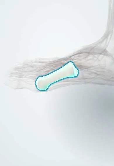
Hallux valgus (bunion) !
Hallux valgus, or bunion, is a deformity where the big toe drifts toward the second toe, leaving a painful bump on the inner side of the foot.
Updated 2026-10-29
Structures involved
Tap one to see what it is and open it in 3D.
- First metatarsal
- Big toe
- Sesamoids
- Adductor hallucis
- Abductor hallucis
- Extensor hallucis longus
- Flexor hallucis longus
- MTP ligaments and plantar plate
- Toe joint capsules
- Second metatarsal
What it is
A forefoot deformity in which the big toe drifts toward the second toe and the first metatarsal drifts toward the midline, leaving a painful bump (bunion) on the inner side of the foot.
Who gets it
Common in adults and more frequent in women; it becomes more common with age. A family history is frequent, especially when it starts in adolescence.
Causes
- Inherited foot shape and ligament laxity
- Narrow, pointed or high-heeled shoes that push the toes together
- Inflammatory joint disease (rheumatoid arthritis) or neuromuscular disease
- Trauma to the first metatarsophalangeal joint
Risk factors
- Female sex
- Family history
- Flatfoot and hypermobility of the first ray
- Generalized joint laxity
- Tight, narrow footwear
- Rheumatoid arthritis
Pathophysiology
Once the big toe starts to drift outward, the tendons that cross the joint pull like a bowstring and push the deformity further. The first metatarsal head slides medially off the sesamoids, the medial capsule stretches and the lateral structures, including the adductor hallucis, tighten. The big toe also rotates, and the inner bump rubs against the shoe.
Classification
- Graded mild, moderate or severe by the hallux valgus angle and the intermetatarsal angle on standing X-rays; cut-offs vary between texts
- Congruent or incongruent joint, judged with the distal metatarsal articular angle
- With or without arthritis of the first metatarsophalangeal joint
Symptoms and signs
- Bump on the inner side of the big toe joint, often red or tender
- Pain on walking or in tight shoes
- Big toe angled toward or under the second toe
- Calluses under the second metatarsal head or on the inner big toe
- Stiffness of the big toe in advanced cases
Diagnosis
- Clinical
- Inspection standing and walking; checks whether the deformity corrects passively, joint motion, first-ray mobility, the second toe, calluses, sensation and pulses.
- Tests
- No lab test is needed; uric acid or inflammatory markers only if gout or inflammatory arthritis is suspected.
- Imaging
- Weight-bearing X-rays of the foot measure the hallux valgus angle, the intermetatarsal angle and the joint congruence and show arthritis.
Differential diagnosis
- Hallux rigidus (arthritis of the first metatarsophalangeal joint)
- Gout of the first metatarsophalangeal joint
- Rheumatoid arthritis of the forefoot
- Bursitis over the first metatarsal head
- Sesamoiditis
Goal of treatment
Relieve pain and allow comfortable walking and footwear; surgery aims to correct the alignment when symptoms persist, not for appearance alone.
Treatment
- Conservative
- Wide, soft shoes with a deep toe box and low heel, bunion pads, toe spacers and orthoses; these ease pain but do not correct the deformity.
- Medical
- Simple analgesics or NSAIDs for flares of pain.
- Surgical
- Osteotomy of the first metatarsal (distal or proximal, chosen by severity) with soft-tissue balancing; fusion of the first tarsometatarsal joint for an unstable first ray; fusion of the big toe joint when there is arthritis or severe deformity.
- Rehabilitation
- After surgery, protected walking in a post-operative shoe, then stretching and gait training.
Complications
- Recurrence of the deformity
- Overcorrection (hallux varus)
- Transfer pain under the lesser metatarsal heads
- Stiffness of the big toe joint
- Nonunion or loss of blood supply to the metatarsal head after osteotomy
- Hammer or crossover second toe
Prognosis
Many people do well with footwear changes. Surgery relieves pain in most patients, though recovery takes months and the deformity can recur.
Prevention
- Shoes with enough width for the forefoot
- Limiting time in narrow, high-heeled shoes
Test yourself
Think of the answer, then tap the question to see it.
Which two radiographic angles are used to grade hallux valgus?
The hallux valgus angle (between the first metatarsal and proximal phalanx) and the intermetatarsal angle (between the first and second metatarsals), on standing X-rays.
Which muscle pulls the big toe laterally and tightens in hallux valgus?
The adductor hallucis, together with the lateral capsule.
Why must foot X-rays for hallux valgus be taken standing?
Weight bearing shows the true alignment; non-weight-bearing films underestimate the angles.
Which footwear factor is most linked to hallux valgus?
Narrow, pointed shoes with high heels, which crowd the toes and load the forefoot.
What is the usual surgical option when hallux valgus is combined with arthritis of the big toe joint?
Fusion (arthrodesis) of the first metatarsophalangeal joint.
Related conditions
- Flatfoot (pes planus)
- Morton neuroma
- Metatarsal fractures (including stress fractures)
- Rheumatoid arthritis
- Gout
Educational information, not medical advice. For any symptom, see a health professional.
Sources: Hallux Valgus (StatPearls (NCBI Bookshelf), 2026).
Spotted a mistake?We are always improving for the community. Tell us what is wrong and our team will review it.Report a mistake
Representative model, for educational purposes only.