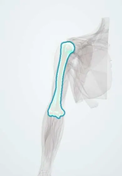
Proximal humerus fracture !
A proximal humerus fracture is a break of the top of the upper arm bone, near the shoulder; it is a typical fracture of older adults with fragile bones.
Updated 2026-10-29
Structures involved
Tap one to see what it is and open it in 3D.
- Humerus
- Humeral head cartilage
- Axillary nerve
- Circumflex humeral arteries
- Supraspinatus
- Subscapularis
- Axillary artery
- Middle deltoid
Why it happens
What it is
A break of the upper end of the humerus: the head, the anatomical or surgical neck, or the greater and lesser tuberosities. It is a typical fragility fracture of older adults.
Who gets it
Very common in older people, especially women with osteoporosis, and rising as the population ages in Colombia and Latin America. In young people it follows high-energy trauma.
Causes
- Fall from standing height onto an outstretched hand or the side of the shoulder
- High-energy trauma (traffic accidents, falls from height) in younger patients
- Fracture-dislocation with a shoulder dislocation; rarely seizures or electric shock
Risk factors
- Older age and female sex
- Osteoporosis
- Frequent falls, poor balance or vision
- Previous fragility fracture
- Long-term corticosteroid use
Pathophysiology
Weak metaphyseal bone fails at the surgical neck, where fractures are most common. The rotator cuff and pectoralis major pull the fragments in set directions: the supraspinatus pulls the greater tuberosity up and back, the subscapularis pulls the lesser tuberosity inward. Damage to the circumflex humeral arteries can cut the blood supply of the head and lead to avascular necrosis.
Classification
- Neer: counts the four parts (head, greater tuberosity, lesser tuberosity, shaft); a part counts as displaced when separated or angulated beyond widely taught thresholds
- One-part (non-displaced), two-, three- and four-part fractures
- Fracture-dislocations and head-splitting fractures
- AO/OTA classification is also used
Symptoms and signs
- Shoulder pain after a fall
- Arm held against the body, unable to lift it
- Swelling and extensive bruising that spreads down the arm and chest over days
- Crepitus with movement
Diagnosis
- Clinical
- Bruising and tenderness over the proximal arm. Clinicians test sensation over the lateral deltoid (regimental badge area) and deltoid contraction for the axillary nerve, plus distal pulses.
- Tests
- Preoperative blood tests as needed; workup for osteoporosis (calcium, vitamin D) after a fragility fracture.
- Imaging
- Shoulder trauma X-ray series including an axillary or scapular Y view to rule out dislocation. CT helps plan surgery in three- and four-part fractures.
Differential diagnosis
- Shoulder dislocation
- Rotator cuff tear
- Clavicle fracture
- Acromioclavicular joint separation
- Pathological fracture through a bone metastasis
Goal of treatment
Achieve a painless, functional shoulder, protect the blood supply of the humeral head and avoid long immobilisation in frail patients.
Treatment
- Conservative
- Most fractures, especially minimally displaced ones, are treated in a sling with early gentle pendulum exercises once pain allows.
- Surgical
- Displaced fractures in active patients may need fixation with a locking plate or intramedullary nail; complex fractures in older adults may get a shoulder replacement (hemiarthroplasty or reverse arthroplasty).
- Rehabilitation
- Physiotherapy progresses from passive to active motion and strengthening to limit stiffness.
- Medical
- Analgesics for pain, and osteoporosis treatment (such as bisphosphonates with calcium and vitamin D) to prevent the next fracture.
Complications
- Shoulder stiffness
- Avascular necrosis of the humeral head
- Malunion or non-union
- Axillary nerve or artery injury
- Fixation failure or screw cut-out in osteoporotic bone
Prognosis
Most minimally displaced fractures heal with a functional shoulder, though some stiffness is common. Results of complex fractures in older people vary, and evidence on surgery versus sling treatment is still debated.
Prevention
- Fall prevention at home
- Diagnosis and treatment of osteoporosis
- Exercise for strength and balance
Test yourself
Think of the answer, then tap the question to see it.
Which nerve is most at risk in a proximal humerus fracture, and how is it tested?
The axillary nerve, which winds around the surgical neck. Clinicians test sensation over the lateral shoulder and deltoid contraction.
What are the four parts of the Neer classification?
The humeral head (articular segment), the greater tuberosity, the lesser tuberosity and the humeral shaft.
Which X-ray view is essential to rule out an associated shoulder dislocation?
An axillary view or a scapular Y view, added to the anteroposterior view.
Why can four-part proximal humerus fractures lead to avascular necrosis?
The fracture can disrupt the circumflex humeral arteries that supply the humeral head.
Related conditions
- Osteoporosis
- Shoulder dislocation
- Rotator cuff tear
- Frozen shoulder (adhesive capsulitis)
- Humeral shaft fracture
- Axillary nerve injury
- Acromioclavicular joint separation
- Clavicle fracture
Educational information, not medical advice. For any symptom, see a health professional.
Sources: Proximal Humerus Fracture (StatPearls (NCBI Bookshelf), 2023).
Spotted a mistake?We are always improving for the community. Tell us what is wrong and our team will review it.Report a mistake
Representative model, for educational purposes only.