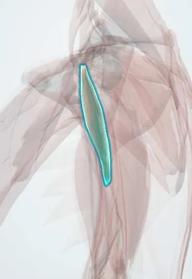
Coracobrachialis !
The coracobrachialis is a slim muscle in the upper inner arm, from the coracoid process to the humerus; it helps raise the arm forward and inward.
Opens in the Durgical app, no sign-up needed.
What it is
A slim muscle from the coracoid to the middle of the humerus.
What it does
Helps raise the arm forward and toward the body.
Key facts
- Origin
- Tip of the coracoid process
- Insertion
- Middle of the medial shaft of the humerus
- Action
- Flexes and adducts the arm
- Nerve
- Musculocutaneous nerve (C5–C7)
- Blood supply
- Brachial artery (muscular branches)
Relations
- Lateral
- Biceps (short head), sharing the conjoint tendon at the tip of the coracoid
- Medial
- Third part of the axillary artery, then the brachial artery, with the median nerve
- Deep
- Subscapularis and the tendons of latissimus dorsi and teres major
- Superficial
- Front deltoid and pectoralis major above; skin of the upper medial arm below
- Contents
- The musculocutaneous nerve passes through it, then runs down between the biceps brachii and the brachialis
How to find it
With the arm abducted and turned out, a narrow ridge just behind the short head of biceps in the upper medial arm tightens when the arm is pulled forward and inward against resistance. The axillary pulse is felt just behind its medial edge.
Examination
- Resisted shoulder flexion with the elbow fully bent (which reduces the biceps' share): weakness, together with weak elbow flexion and numbness of the lateral forearm, points to the musculocutaneous nerve
On imaging
Ultrasound or axial MRI shows it medial to the short head of the biceps below the coracoid, joined to it in the conjoint tendon at the coracoid tip. The musculocutaneous nerve is seen entering it a few centimetres down; ultrasound follows the nerve through it during axillary blocks. Isolated injury is uncommon; MRI shows a strain as oedema.
Clinical notes
- Axillary brachial plexus block: the musculocutaneous nerve often lies inside the coracobrachialis, outside the sheath around the artery, so it is blocked separately
- The musculocutaneous nerve can be trapped within the muscle in weightlifters, with biceps weakness and lateral forearm numbness
- In coracoid transfer (Latarjet) the conjoint tendon moves with the bone, and traction on the musculocutaneous nerve is a known complication
Anatomical variants
- The musculocutaneous nerve sometimes runs past the muscle without piercing it, or travels fused with the median nerve and leaves it lower in the arm; the median nerve may then supply the anterior arm muscles
Development
A derivative of the ventral (flexor) muscle mass, like biceps and brachialis; all three are supplied by the musculocutaneous nerve, which carries anterior-division fibres through the lateral cord.
Test yourself
Think of the answer, then tap the question to see it.
Which nerve pierces the coracobrachialis, and when is it at risk?
The musculocutaneous nerve, a few centimetres below the coracoid; at risk when the conjoint tendon is retracted or moved in a Latarjet.
Why is the musculocutaneous nerve blocked separately in an axillary plexus block?
It often lies inside the coracobrachialis, outside the sheath around the axillary artery.
Which landmarks does the coracobrachialis provide in the arm?
The axillary artery and median nerve lie behind its medial border; its insertion marks mid-arm, where the median nerve crosses the brachial artery.
Educational information, not medical advice. For any symptom, see a health professional.
Its names
- Official Latin (TA2)
- musculus coracobrachialis TA2 2468
- Official Spanish (DeCS)
- Músculo coracobraquial
- English (TA2)
- Coracobrachialis
Related
Sources: Terminologia Anatomica (TA2); Anatomy, Shoulder and Upper Limb, Arm Muscles.
Spotted a mistake?We are always improving for the community. Tell us what is wrong and our team will review it.Report a mistake
Representative model, for educational purposes only.