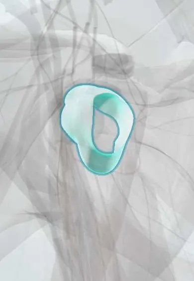
Joints and ligaments · Upper limb
Shoulder capsule !
The shoulder capsule is the loose sleeve that seals the ball-and-socket joint; its looseness lets the shoulder move more than any other joint.
Official name: Shoulder (glenohumeral) joint capsule
Opens in the Durgical app, no sign-up needed.
What it is
The loose bag of tissue around the shoulder ball and socket; its thick parts are the glenohumeral ligaments.
What it does
Seals the joint and holds its fluid; its looseness lets the shoulder move more than any other joint.
Key facts
- Attaches
- Glenoid rim and labrum to the anatomical neck of the humerus (further down medially)
- Function
- Loose for range; reinforced by the glenohumeral ligaments and rotator cuff; axillary pouch below; thickens and shrinks in frozen shoulder; supplied by the axillary and suprascapular nerves and the circumflex humeral and suprascapular arteries
Relations
- Above
- Supraspinatus tendon, which blends with it; above that, the subacromial bursa and the coracoacromial arch
- In front
- Subscapularis tendon and the subscapular recess; the glenohumeral ligaments thicken it from the inside
- Behind
- Infraspinatus tendon and teres minor tendon, which blend with it
- Below
- The axillary recess; below it the axillary nerve and posterior circumflex humeral artery pass back through the quadrangular space
- Contents
- Humeral head and glenoid with the labrum; the long biceps tendon crosses inside the joint but stays outside the synovial membrane
How to find it
It cannot be felt as a structure. The joint line lies below and in front of the acromion; aspiration or injection uses a posterior entry below the posterolateral corner of the acromion, aiming at the coracoid, or an anterior entry lateral to the coracoid.
Examination
- Passive external rotation with the elbow at the side: loss compared with the other side, with normal X-rays, points to frozen shoulder rather than a cuff tear (which keeps passive motion)
- Apprehension and relocation tests: fear in abduction and external rotation that eases when the humeral head is pushed back suggests anterior capsulolabral laxity
- Sulcus sign: downward pull on the arm opens a gap below the acromion, a sign of a lax inferior capsule (multidirectional instability)
On imaging
MR arthrography shows capsular volume, labral tears and capsular tears from the humerus (HAGL). In frozen shoulder MRI shows a thick capsule in the axillary recess and scarring in the rotator interval; ultrasound shows a thick coracohumeral ligament. Joint fluid tracks into the biceps sheath and subscapular recess.
Common problems
- Frozen shoulder (adhesive capsulitis): the capsule becomes inflamed, thick and tight, so the shoulder hurts and stiffens.
- Stretched or torn in a dislocation; a loose capsule lets the shoulder slip.
- Frozen shoulder is more common in people with diabetes and thyroid disease.
How it is diagnosed
Loss of both active and passive motion, especially outward rotation, with normal X-rays. MRI may show a thick capsule.
Treatment
- Gentle stretching, pain control and time: most frozen shoulders improve over months.
- Steroid injection or hydrodilatation in the painful phase.
- Arthroscopic capsular release or manipulation under anesthesia when stiffness persists.
Clinical notes
- Anterior dislocation or its reduction can stretch the axillary nerve: numbness over the lateral deltoid (regimental badge area) and weak abduction, checked before and after reduction
- A stretched capsule in recurrent instability is tightened by capsular shift or arthroscopic plication, usually with labral repair
- Septic arthritis of the shoulder may be subtle in infants and older adults; aspiration of the joint settles the diagnosis
- Frozen shoulder passes through freezing (painful), frozen (stiff) and thawing phases over months; options include physiotherapy, hydrodilatation and capsular release
- Anterior dislocation squares off the shoulder (epaulette sign): the acromion juts out, the rounded deltoid contour is lost and the humeral head is felt below the coracoid
Anatomical variants
- Sublabral foramen and Buford complex (no anterosuperior labrum, with a thick cord-like middle glenohumeral ligament): normal variants that can be mistaken for labral tears on MRI
Development
From week 6 the joint forms between the cartilage models of the humerus and scapula: the centre of the interzone breaks down into the cavity while the mesenchyme around it becomes the capsule; its thicker bands are the glenohumeral ligaments.
Related conditions
- Ehlers–Danlos syndrome
- Polymyalgia rheumatica
- Shoulder dislocation
- Frozen shoulder (adhesive capsulitis)
Test yourself
Think of the answer, then tap the question to see it.
What is the weakest part of the shoulder capsule, and what runs below it?
The inferior capsule (axillary pouch); most dislocations go anterior-inferior through it, and the axillary nerve runs just below.
What is the capsular pattern of the shoulder in frozen shoulder?
External rotation is lost most, then abduction, then internal rotation.
What closes the rotator interval of the shoulder capsule?
Capsule, the coracohumeral ligament and the superior glenohumeral ligament, between the supraspinatus and subscapularis tendons; it thickens early in frozen shoulder.
What is the sulcus sign of the shoulder capsule?
Downward pull on the arm opens a gap below the acromion, a sign of a lax inferior capsule (multidirectional instability).
Educational information, not medical advice. For any symptom, see a health professional.
Its names
- Official Latin (TA2)
- capsula articularis articulationis glenohumeralis TA2 1536
- Official Spanish (DeCS)
- Cápsula articular de la articulación glenohumeral
- English (TA2)
- Shoulder (glenohumeral) joint capsule
- Also called
- shoulder joint capsule
Related
Sources: Terminologia Anatomica (TA2); Anatomy, Shoulder and Upper Limb, Glenohumeral Joint; Anatomy, Shoulder and Upper Limb, Shoulder; 9.7 Development of Joints (OpenStax (Rice University), 2022) · adapted, CC BY 4.0; Anatomy of the Human Body (20th ed.): Development of the Joints (Lea & Febiger (Gray's Anatomy, 1918), 1918) · adapted, Public domain; Anatomía humana (4.ª ed.), tomo 1, cap. 55: Cintura escapular.
Spotted a mistake?We are always improving for the community. Tell us what is wrong and our team will review it.Report a mistake
Representative model, for educational purposes only.