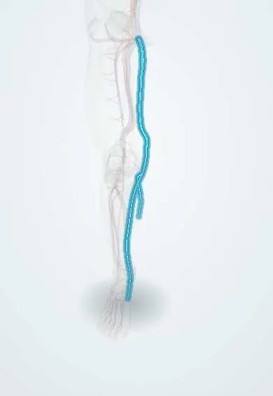
Chronic venous insufficiency !
Chronic venous insufficiency is long-lasting high pressure in the leg veins from faulty valves; it causes swelling, skin changes and sometimes ulcers.
Updated 2026-10-29
Structures involved
Tap one to see what it is and open it in 3D.
- Great saphenous vein
- Small saphenous vein
- Perforating veins
- Leg veins
- Femoral vein
- Popliteal vein
- Top-of-foot veins
- Soleus
- Gastrocnemius (inner head)
In the clinical exam
- Signs of deep vein thrombosis: Homans and the Wells score
- Varicose veins: the Trendelenburg (Brodie–Trendelenburg) test
- Edema: pitting, grading and distribution
In physiology
Why it happens
- Edema and effusions: the four mechanisms
- Pigments in tissue: lipofuscin, hemosiderin, melanin, bilirubin, carbon
- What happens to a thrombus: propagation, embolism, dissolution, organization
- Hyperemia and congestion (the nutmeg liver, heart-failure cells)
- Starling forces at the capillary
- Blood vessels: artery, vein and capillary layers
What it is
Long-standing high pressure in the leg veins (venous hypertension) from faulty valves or blocked veins, causing swelling, skin changes and, in advanced stages, ulcers.
Who gets it
Very common, especially in women, older adults and people who stand for long periods at work; advanced skin changes and ulcers are less frequent but cause long-lasting disability.
Causes
- Primary valve incompetence of the superficial veins
- Post-thrombotic syndrome after deep vein thrombosis
- Venous obstruction (for example iliac vein compression)
- Calf muscle pump failure from immobility or ankle stiffness
- Congenital venous malformations (rare)
Risk factors
- Older age
- Female sex and multiple pregnancies
- Family history
- Obesity
- Prolonged standing or sitting
- Previous deep vein thrombosis
Pathophysiology
Faulty valves or blocked veins mean that calf muscle contraction no longer lowers venous pressure when walking. Persistent venous hypertension pushes fluid, red cells and proteins into the tissues. Inflammation, haemosiderin deposits and fibrosis then damage the skin and subcutaneous fat.
Classification
- CEAP: Clinical, Etiologic, Anatomic and Pathophysiologic
- Clinical classes C0 to C6: C1 telangiectasias, C2 varicose veins, C3 oedema, C4 skin changes, C5 healed ulcer, C6 active ulcer
Symptoms and signs
- Heavy, aching or tired legs, worse at the end of the day and relieved by elevation
- Ankle swelling
- Varicose veins and spider veins
- Itching and night cramps
- Brown pigmentation (haemosiderin), venous eczema
- Lipodermatosclerosis: hard, tight skin above the ankle
- Atrophie blanche (white scar-like areas)
Diagnosis
- Clinical
- History and examination with the patient standing; CEAP clinical class; pulses and the ankle-brachial index to exclude arterial disease.
- Tests
- No blood test confirms it; tests depend on suspected causes or complications.
- Imaging
- Duplex ultrasound is the main test: it shows reflux, which veins are affected and any obstruction or old thrombosis.
Differential diagnosis
- Heart, kidney or liver failure (bilateral oedema)
- Lymphoedema
- Lipoedema
- Drug-induced oedema (for example calcium channel blockers)
- Deep vein thrombosis
Goal of treatment
Lower venous pressure, relieve symptoms, improve skin changes and prevent or heal ulcers.
Treatment
- Conservative
- Graduated compression stockings or bandages, the main treatment, once arterial disease is excluded.
- Lifestyle
- Leg elevation, walking and calf exercises, weight loss and avoiding long periods of standing.
- Medical
- Venoactive drugs (such as flavonoids) for symptoms in some guidelines; emollients and topical steroids for venous eczema.
- Procedure
- Endovenous thermal ablation or sclerotherapy of incompetent superficial veins.
- Surgical
- Vein stripping or ligation in selected cases; stenting of iliac vein obstruction.
Complications
- Venous leg ulcer
- Cellulitis
- Superficial thrombophlebitis
- Bleeding from varicose veins
- Secondary lymphoedema
Prognosis
A chronic, often progressive condition. Compression and treatment of reflux relieve symptoms and lower ulcer recurrence, but adherence over time is the main challenge.
Prevention
- Regular walking and weight control
- Thromboprophylaxis in high-risk patients and early treatment of deep vein thrombosis
- Compression for people with early disease who stand for long periods
Test yourself
Think of the answer, then tap the question to see it.
What does CEAP stand for in chronic venous disease?
Clinical, Etiologic, Anatomic and Pathophysiologic classification.
Which CEAP clinical class is an active venous ulcer?
C6; a healed ulcer is C5 and skin changes are C4.
What is the basic mechanism of chronic venous insufficiency?
Venous hypertension from incompetent valves or obstruction, failing to drain the leg during walking.
What is lipodermatosclerosis?
Inflammatory hardening and fibrosis of the skin and fat above the ankle, giving an inverted champagne-bottle leg.
What is the main imaging test for chronic venous insufficiency?
Venous duplex ultrasound, to show reflux and obstruction.
Related conditions
Educational information, not medical advice. For any symptom, see a health professional.
Sources: Venous Insufficiency (StatPearls (NCBI Bookshelf), 2026).
Spotted a mistake?We are always improving for the community. Tell us what is wrong and our team will review it.Report a mistake
Representative model, for educational purposes only.