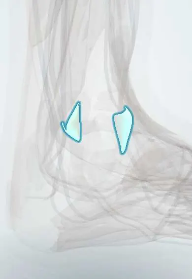
Joints and ligaments · Lower limb
Syndesmosis ligaments !
The syndesmosis ligaments hold the lower ends of the tibia and fibula together just above the ankle; they tear in a high ankle sprain.
Official name: Anterior tibiofibular ligament
Opens in the Durgical app, no sign-up needed.
What it is
A set of ligaments that join the lower ends of the tibia and fibula, just above the ankle joint.
What it does
Keep the two leg bones tightly together so the talus sits snugly between them.
Key facts
- Attaches
- Anterior and posterior inferior tibiofibular, transverse and interosseous ligaments: distal tibia to distal fibula
- Function
- Hold the ankle mortise together; injured in high ankle sprain
- Tight in
- Dorsiflexion and external rotation of foot
Relations
- In front
- Superficial fibular nerve crossing the front of the ankle, the fibularis tertius and toe extensor tendons; the perforating branch of the fibular artery emerges through the interosseous membrane just above
- Behind
- Posterior malleolus of the tibia, where the posterior ligament attaches; behind it, the fibularis longus and brevis tendons and, more medially, the flexor hallucis longus
- Above
- Interosseous membrane, which continues downward as the interosseous ligament
- Below
- Talar dome in the ankle mortise; the transverse ligament deepens the back of the mortise like a lip
How to find it
The anterior inferior tibiofibular ligament runs obliquely just above the ankle joint line, between the front of the tibia and the fibula. Tenderness here, higher than the ATFL, and pain that spreads up the leg suggest a high ankle sprain.
Examination
- Squeeze test: the tibia and fibula are squeezed together at mid-calf; pain felt down at the ankle suggests syndesmosis injury
- External rotation (Kleiger) test: knee at 90°, ankle neutral, foot turned outward; pain over the front of the syndesmosis is positive (medial pain points to the deltoid ligament)
- Fibular translation test: the fibula is moved forward and backward against the tibia; pain or more movement than the other side is positive
- Cotton test: the talus is pushed side to side in the mortise; extra movement suggests syndesmosis and deltoid laxity
On imaging
AP and mortise X-rays about 1 cm above the plafond: tibiofibular clear space (normally under about 6 mm) and overlap; standing or external rotation stress views show subtle widening. X-rays of the whole fibula up to the knee rule out a Maisonneuve fracture. CT (ideally both sides, weight-bearing) shows fibular position; MRI shows the ligaments.
Common problems
- High ankle sprain: the foot is forced to twist outward, often in contact sports. It usually takes longer to heal than a normal sprain.
- Often injured with ankle fractures or deltoid ligament tears.
- If the bones spread apart and stay that way, the ankle can become unstable and wear out.
How it is diagnosed
A physical exam (squeeze test, external rotation test). X-rays, sometimes standing, to look for widening; CT or MRI.
Treatment
- Stable injuries: a boot, limited weight on the foot and physical therapy.
- Unstable injuries: surgery to hold the bones together with a screw or a strong stitch-and-button device.
Clinical notes
- Unstable injuries are fixed with screws or suture-button devices across the syndesmosis; the fibula must sit exactly in the tibial incisura, as malreduction leads to pain and arthritis
- Bony avulsions of the anterior ligament: from the tibia (Tillaux–Chaput tubercle) or from the fibula (Wagstaffe–Le Fort fragment)
- Repeated injury can leave a thickened ligament slip or scar that impinges on the talus at the front of the ankle
Anatomical variants
- The fibular notch of the tibia (incisura) varies in shape on CT, from deep and curved to shallow and flat; a shallow notch makes correct fibular reduction harder to judge
- An accessory lower slip of the anterior inferior tibiofibular ligament (Bassett's ligament) is often present
Development
The distal tibial growth plate closes from medial to lateral in adolescence, so for a while the anterior ligament can pull off the still-open anterolateral corner of the epiphysis (juvenile Tillaux fracture).
Related conditions
Test yourself
Think of the answer, then tap the question to see it.
Which ligaments make up the distal tibiofibular syndesmosis?
The anterior and posterior inferior tibiofibular, transverse and interosseous ligaments.
What mechanism causes a high ankle sprain, and which ligament tears first?
External rotation of the foot (often with dorsiflexion); the anterior inferior tibiofibular ligament tears first.
What is a Maisonneuve injury?
Syndesmosis and deltoid tear with a fracture of the upper fibula, easily missed if only the ankle is X-rayed.
What does a positive squeeze test suggest?
Syndesmosis injury: squeezing the tibia and fibula at mid-calf causes pain down at the ankle.
Educational information, not medical advice. For any symptom, see a health professional.
Its names
- Official Latin (TA2)
- ligamentum tibiofibulare anterius; ligamentum tibiofibulare posterius; ligamentum tibiofibulare transversum; syndesmosis tibiofibularis TA2 1869
- Official Spanish (DeCS)
- Ligamentos tibiofibulares (sindesmosis tibiofibular)
- English (TA2)
- Anterior tibiofibular ligament
- Also called
- inferior tibiofibular joint · ligamentum anterius malleoli lateralis
Related
Sources: Terminologia Anatomica (TA2); Anatomy, Bony Pelvis and Lower Limb: Distal Tibiofibular Joint (Tibiofibular Syndesmosis).
Spotted a mistake?We are always improving for the community. Tell us what is wrong and our team will review it.Report a mistake
Representative model, for educational purposes only.