Joints and ligaments · Lower limb
Interosseous membrane !
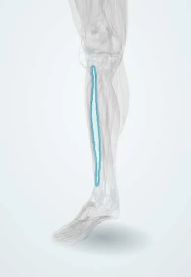
The interosseous membrane is a thin, tough sheet joining the tibia and fibula along the leg; it separates front and back muscles.
Official name: Interosseous membrane of leg
Opens in the Durgical app, no sign-up needed.
What it is
A thin, strong sheet joining the tibia and fibula.
What it does
Ties the two leg bones together and gives attachment to muscles.
Key facts
- Where
- Between interosseous borders of tibia and fibula; gap above for anterior tibial vessels
- Attaches
- Interosseous borders of tibia and fibula; fibers run downward and laterally from tibia to fibula
- Function
- Binds tibia and fibula; origin for anterior and posterior leg muscles; separates anterior and posterior compartments
Relations
- In front
- Tibialis anterior, extensor digitorum longus, extensor hallucis longus and fibularis tertius; the anterior tibial artery and the deep fibular nerve run down on it
- Behind
- Tibialis posterior, which arises from much of it, and flexor hallucis longus lower down
- Above
- Free upper edge just below the proximal tibiofibular joint, with the opening for the anterior tibial vessels
- Below
- Interosseous ligament of the distal tibiofibular syndesmosis
How to find it
Not palpable. In dissection it is reached by lifting the anterior compartment muscles off it from the front or tibialis posterior from behind; on axial images it is the thin line joining the tibia and fibula.
Examination
- Squeeze test: pressing the fibula toward the tibia at mid-calf causes pain at the front of the ankle when the distal syndesmosis and the lower membrane are injured (high ankle sprain)
- External rotation (Kleiger) test: with the knee bent at 90°, turning the foot outward causes anterolateral ankle pain in a syndesmosis injury
On imaging
Axial MRI shows it as a thin low-signal band between the interosseous borders; ultrasound can also follow it. Its tearing is inferred from widening of the tibiofibular clear space at the ankle and from a proximal fibular fracture on radiographs of the whole leg.
Clinical notes
- Maisonneuve injury: syndesmosis and membrane torn up to a proximal fibular fracture, which is why the whole fibula is examined in ankle injuries with a widened mortise
- The anterior tibial artery is tethered where it passes through the upper opening, so it can be injured in proximal tibial fractures, osteotomies and knee dislocations
- In compartment syndrome fasciotomy the crural fascia is opened; the membrane stays intact and limits the anterior and deep posterior compartments
Related conditions
Test yourself
Think of the answer, then tap the question to see it.
What passes through the large opening at the upper end of the interosseous membrane of the leg?
The anterior tibial artery and veins, into the anterior compartment.
What is a Maisonneuve injury?
A tear of the ankle syndesmosis and the interosseous membrane up to a proximal fibular fracture, so the whole fibula is examined in ankle injuries with a widened mortise.
Why is the anterior tibial artery at risk in proximal tibial fractures and knee dislocations?
It is tethered where it passes through the upper opening of the interosseous membrane.
Educational information, not medical advice. For any symptom, see a health professional.
Its names
- Official Latin (TA2)
- membrana interossea cruris TA2 1867
- Official Spanish (DeCS)
- Membrana interósea de la pierna
- English (TA2)
- Interosseous membrane of leg
- Also called
- middle tibiofibular joint
Related
Sources: Terminologia Anatomica (TA2); Anatomy, Bony Pelvis and Lower Limb: Leg Bones.
Spotted a mistake?We are always improving for the community. Tell us what is wrong and our team will review it.Report a mistake
Representative model, for educational purposes only.