Cardiovascular system · Head and neck
Common carotid artery !
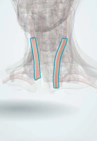
The common carotid artery runs up each side of the neck carrying blood to the head; it splits in two near the top of the voice box.
Official name: Right common carotid artery
Opens in the Durgical app, no sign-up needed.
What it is
The main artery up each side of the neck, in the carotid sheath; its pulse is felt beside the larynx.
What it does
Carries blood to the head; it splits at the level of the upper larynx.
Key facts
- Origin
- Right: brachiocephalic trunk; left: aortic arch
- Branches
- No branches; divides at the upper border of the thyroid cartilage (C3–C4)
- Supplies
- Head and neck
Relations
- Course
- The right one begins behind the right sternoclavicular joint; the left one starts in the thorax from the aortic arch and enters the neck behind the left sternoclavicular joint. Both climb in the carotid sheath beside the trachea and larynx to the upper border of the thyroid cartilage
- Lateral
- Internal jugular vein, in the same sheath, with the vagus nerve in the groove behind and between them
- Medial
- Trachea and oesophagus below, larynx and pharynx above; the lobe of the thyroid gland overlaps it in front, and the recurrent laryngeal nerve runs in the groove between trachea and oesophagus
- Behind
- Sympathetic trunk, longus colli and longus capitis, and the transverse processes of the cervical vertebrae; at about C6 the inferior thyroid artery passes medially behind the sheath, near the middle cervical ganglion, and below C6 the vertebral artery lies deeper; low on the left, the thoracic duct arches behind the sheath
- Superficial
- Sternocleidomastoid, which overlaps it, and the infrahyoid muscles (sternohyoid, sternothyroid, and the omohyoid crossing at about C6); the ansa cervicalis lies on the front of the sheath
How to find it
With the person seated and the head turned slightly toward the side examined, the pulse is felt in the carotid triangle: medial to the front edge of the sternocleidomastoid at the level of the thyroid cartilage, pressing gently backwards toward the transverse processes. One side is examined at a time, and low in the neck rather than at the bifurcation, to avoid pressing on the carotid sinus.
Examination
- Pulse character: a slow-rising, small-volume pulse suggests aortic stenosis; a bounding, collapsing pulse suggests aortic regurgitation
- Auscultation over the bifurcation: a bruit can mean stenosis, but its absence does not exclude a tight narrowing, and a heart murmur can radiate here
- Carotid sinus massage (by clinicians, with ECG monitoring, one side at a time): it can slow some supraventricular tachycardias; a long pause or a marked fall in blood pressure suggests carotid sinus hypersensitivity
On imaging
Duplex ultrasound shows the common carotid, the carotid bulb and both branches, with wall thickness and plaque; CT or MR angiography maps the whole course from the aortic arch to the skull base.
Clinical notes
- Carotid sinus hypersensitivity: in older adults, turning the head, a tight collar or shaving can trigger a reflex slowing of the heart, a fall in blood pressure and fainting
- Central venous access: the internal jugular vein lies lateral to the palpable carotid; ultrasound guidance reduces accidental puncture of the artery
- A carotid body tumour (paraganglioma) at the bifurcation splays the internal and external carotid arteries apart (lyre sign) and moves side to side but not up and down
- Penetrating neck injuries are described by zones; zone II (cricoid cartilage to angle of the mandible) holds most of the common carotid and is the easiest to explore
- The omohyoid crossing divides it: below, it lies deep under the sternocleidomastoid and infrahyoid muscles; above, in the carotid triangle, it is superficial and is where it is usually exposed or tied
Anatomical variants
- A shared origin of the brachiocephalic trunk and the left common carotid artery (the so-called bovine arch) is the commonest aortic arch variant
- The bifurcation level varies: it can lie as high as the hyoid bone or below the thyroid cartilage, which matters for surgery and ultrasound
Development
The common carotid and the first part of the internal carotid come from the third pharyngeal arch arteries.
Memory aid
Carotid sheath contents: "I See 10 CC's in the IV" (Internal Carotid above, cranial nerve 10 = vagus, Common Carotid, Internal jugular Vein).
Related conditions
Test yourself
Think of the answer, then tap the question to see it.
What are the origins of the right and left common carotid arteries?
Right: the brachiocephalic trunk, behind the right sternoclavicular joint. Left: the aortic arch, in the thorax.
How are the contents of the carotid sheath arranged around the common carotid artery?
Common carotid medially, internal jugular vein laterally, vagus nerve behind and between them; the sympathetic trunk is behind the sheath, not in it.
At what level does the common carotid artery divide?
At the upper border of the thyroid cartilage (C3–C4); it has no branches before that.
Why is the carotid pulse felt one side at a time and low in the neck?
Pressing both can cut blood flow to the brain, and pressure on the carotid sinus can slow the heart and drop the blood pressure.
Against which bony landmark can the common carotid artery be pressed?
The carotid tubercle (anterior tubercle of C6), where the omohyoid crosses the carotid sheath.
Educational information, not medical advice. For any symptom, see a health professional.
Its names
- Official Latin (TA2)
- carotis communis dextra TA2 4181
- Official Spanish (DeCS)
- Arteria Carótida Común DeCS D017536
- English (TA2)
- Right common carotid artery
- Also called
- Carótida primitiva
Related
Sources: Terminologia Anatomica (TA2); DeCS; Anatomy, Head and Neck, Anterior: Common Carotid Arteries; Anatomy, Head and Neck: Carotid Arteries; Tratado de anatomía humana (9.ª ed.), tomo II, Libro IV, sección 2 Arterias, caps. I-III (sistema de la arteria pulmonar; aorta y ramas del cayado hasta la carótida interna).
Spotted a mistake?We are always improving for the community. Tell us what is wrong and our team will review it.Report a mistake
Representative model, for educational purposes only.