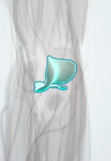
Elbow cartilage (humerus) !
The elbow cartilage of the humerus covers the lower end of the upper arm bone; the radius and ulna glide on it as the elbow bends.
Official name: Cartilage of the capitulum and trochlea
Opens in the Durgical app, no sign-up needed.
What it is
The smooth covering of the lower end of the humerus, where the elbow bends.
What it does
Lets the radius and ulna glide on the humerus.
Key facts
- Where
- Capitulum (for the radial head) and trochlea (for the ulna)
- Function
- Hyaline cartilage; smooth, low-friction joint surface; wears in osteoarthritis
Relations
- Below
- Ulnar notch cartilage on the trochlea medially and radial head cartilage on the capitulum laterally
- In front
- Coronoid fossa and radial fossa just above, which receive the coronoid process and the radial head in full flexion; then the elbow capsule and brachialis
- Behind
- Olecranon fossa above the trochlea, which takes the olecranon tip in extension; the triceps tendon behind
- Medial
- Medial epicondyle with the UCL (elbow) origin; the ulnar nerve runs behind it
- Lateral
- Lateral epicondyle with the RCL (elbow) and the common extensor tendon
How to find it
With the elbow extended, a small hollow at the back, just below the lateral epicondyle, overlies the groove between the capitulum and the radial head; the trochlea cannot be felt. Both surfaces are exposed from behind, through an olecranon osteotomy or a triceps-sparing approach.
On imaging
AP and lateral X-rays; on the lateral view a 'double-arc' sign marks a displaced capitulum shear fracture. MRI or MR arthrography stages osteochondritis dissecans. A notch at the back of the capitulum, where its cartilage ends next to the lateral epicondyle, is a normal pseudodefect, not a lesion.
Clinical notes
- Capitulum shear fractures (Hahn–Steinthal with a large bony piece, Kocher–Lorenz mostly cartilage) follow a fall on the outstretched hand; the fragment flips up in front of the humerus
- Unstable osteochondritis dissecans fragments come loose as loose bodies that catch or lock the elbow; options range from rest from throwing to drilling, fixation or osteochondral grafting
- Intra-articular distal humerus fractures need exact restoration of the trochlea and capitulum; an olecranon osteotomy is a common way to expose them from behind
Development
Elbow ossification centres appear in a fixed order, roughly every two years from about age 1 (ages vary between texts): capitulum, radial head, medial epicondyle, trochlea, olecranon, lateral epicondyle. The trochlea may ossify from several pieces, a normal look that mimics a fracture.
Memory aid
CRITOE (or CRITOL) gives the order of the elbow ossification centres: Capitulum, Radial head, Internal (medial) epicondyle, Trochlea, Olecranon, External (lateral) epicondyle.
Test yourself
Think of the answer, then tap the question to see it.
Which bones articulate with the two surfaces of the distal humeral cartilage?
The trochlea with the trochlear notch of the ulna and the capitulum with the radial head.
Who typically gets osteochondritis dissecans of the capitulum, and why?
Adolescent throwers and gymnasts, from repeated valgus compression of the radial head on the capitulum.
On a child's lateral elbow X-ray, what should the anterior humeral line cross?
The middle third of the capitulum; if it passes in front, an extension supracondylar fracture is suspected.
Educational information, not medical advice. For any symptom, see a health professional.
Its names
- Official Latin (TA2)
- cartilago articularis capituli et trochleae humeri TA2 382
- Official Spanish (DeCS)
- Cartílago articular del capítulo y la tróclea del húmero
- English (TA2)
- Cartilage of the capitulum and trochlea
Related
Sources: Terminologia Anatomica (TA2); Anatomy, Shoulder and Upper Limb, Elbow Joint; Anatomy of the Human Body (20th ed.): Surface Anatomy of the Upper Extremity (Lea & Febiger (Gray's Anatomy, 1918), 1918) · adapted, Public domain.
Spotted a mistake?We are always improving for the community. Tell us what is wrong and our team will review it.Report a mistake
Representative model, for educational purposes only.