Joints and ligaments · Upper limb
UCL (elbow) !
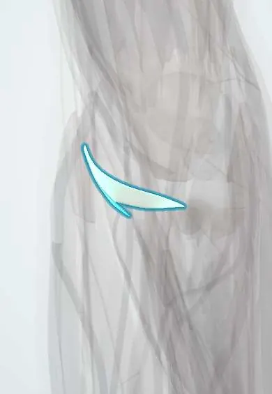
The ulnar collateral ligament is on the inner side of the elbow; it stops the joint opening inward and takes heavy strain in throwing.
Official name: Ulnar (medial) collateral ligament of the elbow
Opens in the Durgical app, no sign-up needed.
What it is
A fan of strong bands on the inner side of the elbow, from the medial epicondyle to the ulna.
What it does
Stops the elbow opening on the inner side; it takes huge stress when throwing.
Key facts
- Attaches
- Medial epicondyle to the sublime tubercle of the coronoid (anterior bundle), olecranon (posterior bundle), transverse band (some texts: anterior, middle and posterior bundles; transverse band = Cooper's ligament)
- Function
- Main restraint to valgus stress (throwing)
- Tight in
- Anterior bundle throughout (front band in extension, back band in flexion); posterior bundle >90° flexion
Relations
- Course
- From the front-lower surface of the medial epicondyle it fans down to the sublime tubercle of the coronoid (anterior bundle) and the medial olecranon (posterior bundle); the transverse band joins the two on the ulna
- Superficial
- Flexor-pronator mass and the common flexor tendon (flexor digitorum superficialis and flexor carpi ulnaris over the anterior bundle), which help resist valgus
- Behind
- Ulnar nerve in the cubital tunnel, lying on the posterior bundle
- Deep
- Medial capsule and the medial side of the trochlea
How to find it
With the elbow slightly bent, the anterior bundle is felt as a firm band from the front-lower medial epicondyle down to the sublime tubercle, in front of the ulnar nerve. Tenderness along this band in a thrower suggests the ligament; tenderness higher, on the epicondyle itself, suggests the flexor origin.
Examination
- Valgus stress test: elbow flexed about 20–30° to unlock the olecranon, humerus steadied; medial pain or more opening than the other elbow suggests a UCL injury
- Moving valgus stress test: shoulder abducted and externally rotated, valgus kept on the elbow while it is extended quickly from full flexion; medial pain reproduced between about 120° and 70° is positive
- Milking manoeuvre: the thumb is pulled outward with the forearm supinated and the elbow flexed past 90°, loading the anterior bundle; medial pain or apprehension is positive
On imaging
MR arthrography shows full and partial tears; the 'T sign' is contrast tracking down along the sublime tubercle under an undersurface tear. Stress ultrasound or stress X-rays measure medial opening against the other elbow. X-rays show calcification within the ligament and posteromedial olecranon osteophytes.
Common problems
- Sprain or tear from repeated throwing (baseball pitchers, javelin), when the elbow is forced open on the inner side.
- Inner elbow pain and loss of throwing speed; sometimes a 'pop'.
- Can tear with an elbow dislocation.
How it is diagnosed
Pain with valgus stress tests (for example the moving valgus test). MRI or MR arthrogram shows the ligament; stress ultrasound can show gapping.
Treatment
- Rest from throwing, physical therapy and a gradual throwing program for partial injuries.
- UCL reconstruction ('Tommy John' surgery) with a tendon graft for complete tears in throwers.
- UCL repair with internal bracing in selected younger patients.
Clinical notes
- UCL reconstruction (popularly 'Tommy John surgery') replaces the anterior bundle with a tendon graft, often the palmaris longus, passed through tunnels in the medial epicondyle and sublime tubercle; return to throwing usually takes about a year
- Valgus extension overload: chronic laxity drives the olecranon against the medial olecranon fossa, forming posteromedial osteophytes and loose bodies
- The ulnar nerve and the medial forearm skin nerve are at risk in medial approaches; the ulnar nerve is often moved forward (transposed) during reconstruction
- Often injured in elbow dislocations, but last: soft-tissue failure spreads from lateral to medial (Horii circle), and the anterior bundle may survive
Development
The medial epicondyle ossifies from its own centre (the 'I' of CRITOE) and fuses late; after an elbow dislocation in a child, an avulsed epicondyle can be trapped in the joint and mistaken for the trochlear centre.
Related conditions
- Cubital tunnel syndrome (ulnar neuropathy at the elbow)
- Medial epicondylitis (golfer's elbow)
- Elbow dislocation
Test yourself
Think of the answer, then tap the question to see it.
Which part of the elbow's ulnar collateral ligament is the primary restraint to valgus stress?
The anterior bundle, from the medial epicondyle to the sublime tubercle of the coronoid; it resists valgus from about 20° to 120° of flexion, where throwing loads peak.
In which phases of throwing is the elbow's ulnar collateral ligament injured?
Late cocking and early acceleration, when valgus torque at the elbow is highest.
Why does ulnar neuritis often accompany ulnar collateral ligament injury of the elbow in throwers?
The posterior bundle forms the floor of the cubital tunnel, so the ulnar nerve lies directly on it.
What is the moving valgus stress test for the elbow's ulnar collateral ligament?
Valgus is kept on the elbow while it is extended quickly from full flexion, shoulder abducted and externally rotated; medial pain between about 120° and 70° is positive.
What structure fails instead of the elbow's ulnar collateral ligament when children throw repeatedly?
The medial epicondyle growth plate, which can avulse.
Educational information, not medical advice. For any symptom, see a health professional.
Its names
- Official Latin (TA2)
- ligamentum collaterale ulnare cubiti TA2 1776
- Official Spanish (DeCS)
- Ligamento colateral ulnar del codo
- English (TA2)
- Ulnar (medial) collateral ligament of the elbow
- Also called
- medial collateral ligament of elbow · UCL
Related
Sources: Terminologia Anatomica (TA2); Anatomy, Shoulder and Upper Limb, Elbow Collateral Ligaments.
Spotted a mistake?We are always improving for the community. Tell us what is wrong and our team will review it.Report a mistake
Representative model, for educational purposes only.