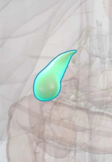
Gallbladder cancer !
Gallbladder cancer is a malignant tumour of the gallbladder lining; early on it rarely causes symptoms, so it is often found late or after gallbladder removal.
Updated 2026-10-29
Structures involved
Tap one to see what it is and open it in 3D.
- Gallbladder
- Cystic duct
- Common bile duct
- Liver
- Liver segment IV
- Liver segment V
- Common hepatic duct
- Celiac and gastric lymph nodes
- Portal vein
- Proper hepatic artery
Why it happens
- Precancerous conditions: when chronic inflammation, metaplasia or hyperplasia raise the risk
- Gallbladder and bile ducts
What it is
Malignant tumour arising from the lining of the gallbladder, usually an adenocarcinoma. Early disease rarely causes symptoms, so it is often found late or by chance after a cholecystectomy.
Who gets it
Uncommon worldwide but with striking geographic variation: rates are among the highest in Chile and the Andean region of South America, and in northern India. It is more frequent in women and older adults, following the pattern of gallstones, so it matters in Latin American practice.
Causes
- Chronic gallbladder inflammation, most often from long-standing gallstones
- Porcelain gallbladder (wall calcified by chronic inflammation)
- Gallbladder polyps, especially larger or growing ones
- Anomalous pancreaticobiliary duct junction, which lets pancreatic juice reflux into the bile ducts
- Chronic typhoid (Salmonella Typhi) carriage
Risk factors
- Gallstones, especially large or long-standing ones
- Female sex and older age
- Andean or Indigenous American ancestry and living in high-incidence areas
- Obesity
- Porcelain gallbladder and gallbladder polyps
- Choledochal cysts and primary sclerosing cholangitis
Pathophysiology
Chronic irritation of the mucosa by stones and bile drives a sequence from metaplasia through dysplasia to carcinoma. The gallbladder wall is thin, has no submucosa and lies directly on the liver, so the tumour spreads early into the liver bed (segments IV and V), the lymph nodes and the peritoneum. Invasion of the bile ducts causes obstructive jaundice.
Classification
- TNM staging: the depth of wall invasion (T stage) is the key prognostic factor
- Incidental (found in the cholecystectomy specimen) or clinically suspected
- Histology: adenocarcinoma in the large majority; squamous and adenosquamous types are rare
Symptoms and signs
- Often none in early stages
- Right upper quadrant pain that may mimic biliary colic or cholecystitis
- Painless obstructive jaundice with itching, dark urine and pale stools in advanced disease
- Weight loss, loss of appetite and fatigue
- Palpable mass in the right upper quadrant
- Nausea and vomiting from duodenal obstruction in late disease
Diagnosis
- Clinical
- Suspected in older patients with gallstones who develop jaundice, weight loss or a right upper quadrant mass. Many cases are found by the pathologist after a routine cholecystectomy.
- Tests
- Liver function tests with a cholestatic pattern (raised bilirubin and alkaline phosphatase). Tumour markers such as CA 19-9 and CEA may be raised but are not diagnostic. Histology confirms it; percutaneous biopsy is often avoided when the tumour looks resectable, to limit seeding.
- Imaging
- Ultrasound comes first: wall thickening, a polypoid mass or a mass replacing the gallbladder. Contrast CT and MRI with MRCP stage the tumour (liver invasion, bile ducts, vessels and nodes); PET-CT is sometimes added.
Differential diagnosis
- Acute or chronic cholecystitis
- Xanthogranulomatous cholecystitis
- Adenomyomatosis or a benign gallbladder polyp
- Cholangiocarcinoma
- Liver tumour or metastasis invading the gallbladder
- Pancreatic head cancer (obstructive jaundice)
Goal of treatment
Complete removal with clear margins when the disease is localised; otherwise relief of jaundice and symptoms, systemic therapy to prolong life, and good palliative care.
Treatment
- Surgical
- Simple cholecystectomy can be enough for very early tumours confined to the mucosa (T1a).
- Surgical
- Extended (radical) cholecystectomy for more invasive tumours: the gallbladder is removed with a margin of the liver bed (segments IVb and V) and the regional lymph nodes, sometimes with the bile duct. Incidental cancers beyond T1a usually go back to surgery.
- Medical
- Systemic chemotherapy (antimetabolite and platinum combinations), sometimes with immune checkpoint inhibitors, after surgery or for advanced disease; regimens vary between guidelines.
- Procedure
- Palliative biliary drainage with an endoscopic or percutaneous stent to relieve jaundice and itching; a duodenal stent for gastric outlet obstruction.
- Conservative
- Palliative care for pain, nutrition and symptom control.
Complications
- Obstructive jaundice and cholangitis
- Gastric outlet or duodenal obstruction
- Liver invasion and liver failure
- Peritoneal carcinomatosis and malignant ascites
- Port-site or peritoneal seeding after surgery for an unsuspected cancer
Prognosis
Prognosis depends mainly on the stage. Early tumours found incidentally can be cured, but most patients present with advanced disease and survival is poor.
Prevention
- Cholecystectomy for symptomatic gallstones
- Cholecystectomy is often advised for porcelain gallbladder and for larger or growing polyps; size thresholds vary between guidelines
- Pathological examination of every cholecystectomy specimen
Test yourself
Think of the answer, then tap the question to see it.
What is the strongest risk factor for gallbladder cancer?
Gallstones with chronic inflammation, especially large, long-standing stones.
Which liver segments are removed in an extended cholecystectomy?
Segments IVb and V (the gallbladder bed), together with the regional lymph nodes.
Why does gallbladder cancer invade the liver early?
The gallbladder wall is thin, has no submucosa, and its bed lies directly on the liver (segments IV and V).
What is a porcelain gallbladder and why does it matter?
A gallbladder wall calcified by chronic inflammation; it is linked to cancer (less strongly than once thought), and cholecystectomy is often advised.
In which parts of the world is gallbladder cancer especially common?
Chile and the Andean region of South America, and northern India, mostly in women.
Related conditions
- Gallstones (cholelithiasis)
- Acute cholecystitis
- Choledocholithiasis
- Acute cholangitis
- Obesity
- Pancreatic cancer
Educational information, not medical advice. For any symptom, see a health professional.
Sources: Gallbladder Carcinoma (StatPearls (NCBI Bookshelf), 2024).
Spotted a mistake?We are always improving for the community. Tell us what is wrong and our team will review it.Report a mistake
Representative model, for educational purposes only.