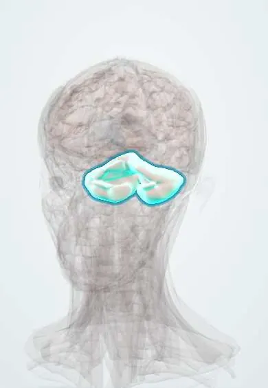
Childhood brain tumours (medulloblastoma, pilocytic astrocytoma, craniopharyngioma) !
Brain tumours are the most common solid cancers in children. Most grow near the cerebellum and often show up as morning headache and vomiting.
Updated 2026-11-01
Structures involved
Tap one to see what it is and open it in 3D.
- Cerebellum
- Vermis
- Fourth ventricle
- Pituitary gland
- Sella turcica
- Optic chiasm
- Hypothalamus
- Third ventricle
- Lateral ventricles
- Pons
- Cerebellar peduncles
- Pineal gland
Why it happens
- Hereditary cancer syndromes: the table to know
- Tumours of infancy and childhood
- Glial cells: astrocytes, oligodendrocytes, microglia, Schwann cells
What it is
Primary tumours of the central nervous system in children. Unlike adults, most are infratentorial (posterior fossa), and the classic exam trio is pilocytic astrocytoma, medulloblastoma and craniopharyngioma.
Who gets it
Brain tumours are the most common solid tumours and the second most common cancer in children after leukaemia, and a leading cause of cancer death in childhood. In Colombia they are part of the childhood cancers under mandatory notification to the national public health surveillance system, and delays in diagnosis are common because early symptoms are nonspecific.
Causes
- Mostly unknown
- Genetic syndromes: neurofibromatosis type 1 (optic pathway gliomas), tuberous sclerosis (subependymal giant cell astrocytoma), Li-Fraumeni, Gorlin and Turcot syndromes (medulloblastoma)
- Previous radiation to the head
Risk factors
- Hereditary cancer predisposition syndromes
- Previous cranial radiotherapy
- Family history of brain tumours
Pathophysiology
Posterior fossa tumours grow in or next to the cerebellum and fourth ventricle, block cerebrospinal fluid flow and cause obstructive hydrocephalus and raised intracranial pressure, plus cerebellar signs. Medulloblastoma is a malignant embryonal tumour of the cerebellar vermis that can seed through the cerebrospinal fluid ("drop metastases" to the spinal cord). Pilocytic astrocytoma is a low-grade, usually cystic cerebellar tumour. Craniopharyngioma arises from remnants of Rathke pouch above the sella turcica and compresses the optic chiasm, pituitary gland and hypothalamus.
Classification
- Pilocytic astrocytoma: most common, low grade (WHO grade 1); cerebellum, cystic with a mural nodule; Rosenthal fibres; often associated with NF1 when in the optic pathway
- Medulloblastoma: most common malignant brain tumour in children; cerebellar vermis; small round blue cells with Homer Wright rosettes; molecular groups (WNT, SHH, group 3, group 4)
- Craniopharyngioma: suprasellar, benign but locally aggressive; calcified and cystic with "motor oil" fluid; derived from Rathke pouch
- Ependymoma: fourth ventricle; perivascular pseudorosettes; causes hydrocephalus
- Diffuse midline glioma (brainstem glioma): pons; poor prognosis
Symptoms and signs
- Morning headache with vomiting, often relieved by vomiting
- Gait and balance problems (truncal ataxia with vermis tumours; limb ataxia with hemisphere tumours)
- Increasing head size and bulging fontanelle in infants
- Double vision or head tilt from abducens palsy
- Bitemporal hemianopia and reduced vision (craniopharyngioma)
- Short stature, delayed puberty, excessive thirst and urination (hypopituitarism, diabetes insipidus)
- Irritability, school decline or developmental regression
Diagnosis
- Clinical
- Full neurological examination with fundoscopy, visual fields, growth and puberty assessment and head circumference in infants. Persistent headache with vomiting or neurological signs needs urgent imaging.
- Tests
- Histology and molecular profiling of the resected tumour. Lumbar cerebrospinal fluid cytology (after surgery) for medulloblastoma staging. Pituitary hormone and sodium tests for sellar tumours. Formal eye and hearing assessment.
- Imaging
- MRI of the brain and whole spine with contrast is the test of choice. Pilocytic astrocytoma: cyst with an enhancing mural nodule. Medulloblastoma: solid midline vermis mass filling the fourth ventricle. Craniopharyngioma: suprasellar mass with calcifications, best seen on CT.
Differential diagnosis
- Migraine and other primary headaches
- Idiopathic intracranial hypertension
- Hydrocephalus from other causes
- Brain abscess or neurocysticercosis
- Pituitary adenoma and Rathke cleft cyst (sellar masses)
- Gastroenteritis or other causes of recurrent vomiting
Goal of treatment
Relieve hydrocephalus and raised pressure, remove as much tumour as safely possible, give risk-adapted radiotherapy and chemotherapy, and limit long-term effects on growth, hormones, hearing and learning.
Treatment
- Acute
- Corticosteroids for edema and urgent treatment of hydrocephalus (external ventricular drain, endoscopic third ventriculostomy or shunt).
- Surgical
- Maximal safe surgical resection: often curative alone for pilocytic astrocytoma; for craniopharyngioma surgery is balanced against hypothalamic injury.
- Procedure
- Craniospinal radiotherapy with a posterior fossa boost for medulloblastoma in children over about 3 years; focal radiotherapy for residual craniopharyngioma. Proton therapy reduces long-term damage where available.
- Medical
- Chemotherapy for medulloblastoma and for low-grade gliomas that cannot be removed; targeted therapy (e.g. BRAF and MEK inhibitors) for some gliomas. Hormone replacement (including desmopressin) for pituitary insufficiency.
- Follow-up
- Neurological and vital-sign checks for raised intracranial pressure, fluid balance and sodium after sellar surgery, and long-term follow-up of growth, puberty, hearing, vision and school performance.
Complications
- Obstructive hydrocephalus
- Posterior fossa mutism syndrome after surgery
- Panhypopituitarism, diabetes insipidus and hypothalamic obesity (craniopharyngioma)
- Hearing loss, learning difficulties and growth failure after treatment
- Recurrence and second cancers after radiotherapy
Prognosis
About three in four children with a brain tumour survive at least five years. Pilocytic astrocytoma has an excellent prognosis after complete resection; medulloblastoma survival depends on age, spread and molecular group (WNT has the best outcome); diffuse midline glioma has a very poor prognosis.
Test yourself
Think of the answer, then tap the question to see it.
Where are most brain tumours located in children, compared with adults?
Infratentorial (posterior fossa) in children; supratentorial in adults.
What is the classic MRI appearance of pilocytic astrocytoma?
A cystic cerebellar mass with an enhancing mural nodule.
Which childhood tumour arises in the cerebellar vermis and can spread through cerebrospinal fluid to the spine?
Medulloblastoma ("drop metastases").
A child has bitemporal hemianopia, short stature and a calcified suprasellar mass. What is the likely tumour?
Craniopharyngioma, derived from Rathke pouch remnants.
Which histological finding is typical of pilocytic astrocytoma?
Rosenthal fibres (eosinophilic corkscrew fibres).
Related conditions
- Hydrocephalus
- Raised intracranial pressure and brain herniation
- Neurofibromatosis (types 1 and 2)
- Tuberous sclerosis complex
- Hypopituitarism (incl. Sheehan syndrome and pituitary apoplexy)
- Diabetes insipidus (central and nephrogenic)
- Pituitary adenoma
- Glioblastoma
- Retinoblastoma
- Neuroblastoma
- Brain metastases
Educational information, not medical advice. For any symptom, see a health professional.
Sources: Brain tumor - children: MedlinePlus Medical Encyclopedia (MedlinePlus (U.S. National Library of Medicine), 2026); Childhood Medulloblastoma and Other Central Nervous System Embryonal Tumors Treatment (PDQ®)–Patient Version (National Cancer Institute (NCI), 2026).
Spotted a mistake?We are always improving for the community. Tell us what is wrong and our team will review it.Report a mistake
Representative model, for educational purposes only.