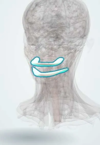
Digestive system · Head and neck
Gums (gingiva) !
The gums (gingiva) are the pink tissue covering the jawbones and sealing around each tooth; inflamed gums can lead to bone loss around the teeth.
Opens in the Durgical app, no sign-up needed.
What it is
The pink tissue that covers the tooth-bearing bone of the jaws and fits tightly around the neck of each tooth.
What it does
Protects the bone and the roots, and seals the tooth sockets against germs.
Key facts
- Where
- Mucosa covering the alveolar processes and the necks of the teeth; attached gingiva bound to bone, free margin around each tooth
- Blood supply
- Branches of the maxillary artery (alveolar, palatine, buccal) and, for the lower gums, the inferior alveolar, lingual and facial arteries
- Nerve
- V2 (superior alveolar, greater palatine, nasopalatine nerves) for the upper gums; V3 (inferior alveolar, buccal, lingual nerves) for the lower gums
- Function
- Seals around the teeth and protects the bone; inflamed in gingivitis and lost with bone in periodontitis
Relations
- Course
- A continuous band along both jaws on the outer (vestibular) side and the inner side, blending with the palatal mucosa above and the floor of the mouth below
- Deep
- Alveolar bone of the maxilla and mandible and the periodontal ligament; at the tooth, the junctional epithelium attaches to the enamel near the cementoenamel junction
How to find it
Seen at the necks of the teeth: firm, pale pink attached gingiva, the scalloped free margin and the papillae filling the spaces between teeth. The mucogingival junction is the line where it meets the redder, movable alveolar mucosa.
Examination
- Periodontal probing: a thin probe slid into the sulcus measures its depth and shows bleeding; a healthy sulcus is shallow (up to about 3 mm), while deeper pockets with attachment loss point to periodontitis
On imaging
The gum itself does not show on radiographs; it is assessed by looking and probing. Bitewing, periapical or panoramic radiographs show what lies under it: the alveolar bone crest, normally just below the enamel-cementum junction. Horizontal or vertical bone loss and calculus point to periodontitis; in gingivitis alone the bone is normal. Cone beam CT is kept for implant or surgical planning.
Common problems
- Gingivitis: red, swollen gums that bleed when brushing.
- Periodontitis: the gum and the bone recede and the teeth loosen.
Clinical notes
- Gum overgrowth occurs in pregnancy, with an exaggerated response to plaque and sometimes a localised pregnancy epulis (pyogenic granuloma)
- Drug-induced gum overgrowth is classically caused by phenytoin, calcium channel blockers such as nifedipine, and ciclosporin
- Bleeding gums can also reflect a general disorder such as leukaemia, a low platelet count or scurvy (vitamin C deficiency)
- Necrotising gingivitis (painful, ulcerated papillae and foul breath) appears with stress, smoking and weakened immunity
Development
Its epithelium comes from the ectoderm lining the primitive mouth; the junctional epithelium that seals the gum to the tooth first forms from the reduced enamel epithelium as the tooth erupts.
Related conditions
- I-cell disease (mucolipidosis II)
- Scurvy (vitamin C deficiency)
- Periodontitis
- Stevens–Johnson syndrome and toxic epidermal necrolysis
- Pemphigus vulgaris
- Lichen planus
- Kaposi sarcoma
- Mucous membrane (cicatricial) pemphigoid
- Von Willebrand disease
- Inherited platelet function disorders (Glanzmann thrombasthenia, Bernard–Soulier syndrome)
- Recurrent aphthous stomatitis
- Leukocyte adhesion deficiency
Test yourself
Think of the answer, then tap the question to see it.
What is the difference between gingivitis and periodontitis?
Gingivitis is reversible inflammation of the gum alone; periodontitis adds loss of periodontal ligament and bone (attachment loss) and does not reverse.
What is the earliest common sign of gum inflammation?
Bleeding gums on brushing or probing.
What does periodontal probing of the gingival sulcus show?
Its depth and any bleeding: a healthy sulcus is shallow (up to about 3 mm); deeper pockets with attachment loss point to periodontitis.
Which drugs classically cause gum overgrowth?
Phenytoin, calcium channel blockers such as nifedipine, and ciclosporin.
Educational information, not medical advice. For any symptom, see a health professional.
Sources: Anatomy, Head and Neck, Oral Gingiva; Histology, Oral Mucosa.
Spotted a mistake?We are always improving for the community. Tell us what is wrong and our team will review it.Report a mistake
Representative model, for educational purposes only.