Conditions · Ophthalmology and ENT
Recurrent aphthous stomatitis !
Repeated painful, round mouth ulcers (canker sores) that heal on their own in one to two weeks. They are common and not contagious.
Updated 2026-11-01
Structures involved
Tap one to see what it is and open it in 3D.
- Tongue
- Gums (gingiva)
- Soft palate
- Palatine tonsils
- Pharynx
- Lingual nerve
- Sublingual gland
- Neck lymph nodes
- Mandible
Why it happens
- Patterns of acute inflammation: serous, fibrinous, purulent, abscess, ulcer
- Oral cavity, tongue and teeth
What it is
The most common cause of recurrent oral ulcers. Aphthous ulcers are round or oval, shallow, painful ulcers with a yellow-grey fibrinous base and a red halo, found on the non-keratinised mucosa of the mouth (inner lips, cheeks, floor of the mouth, sides and underside of the tongue, soft palate). They come back at intervals in otherwise healthy people. The same type of ulcer can also be a sign of a systemic disease, which is then called complex aphthosis or aphthous-like ulcers.
Who gets it
It affects roughly one in five people at some point, with wide variation between populations. It usually begins in childhood or adolescence, is somewhat more frequent in women and tends to become milder after middle age. A family history is common. It is a frequent reason for consultation in general dentistry and primary care in Colombia and Latin America.
Causes
- Cause not fully known; a dysregulated local immune response against the oral mucosa in genetically predisposed people
- Local trauma: biting, dental work, rough toothbrushing, orthodontic appliances
- Deficiencies of iron, vitamin B12 or folate
- Emotional stress and lack of sleep
- Hormonal changes, for example around menstruation
- Some foods and toothpastes containing sodium lauryl sulphate; stopping smoking can unmask ulcers
- Systemic disease: Behçet disease, coeliac disease, Crohn disease and ulcerative colitis, HIV infection, cyclic neutropenia and periodic fever syndromes (PFAPA)
- Drugs such as NSAIDs, nicorandil and some chemotherapy agents (aphthous-like ulcers)
Risk factors
- Family history of mouth ulcers
- Female sex and younger age
- Haematinic deficiency (iron, B12, folate)
- Stress and local mucosal trauma
- Coeliac disease or inflammatory bowel disease
- Immunosuppression, including HIV infection
Pathophysiology
A trigger such as minor trauma activates a T-cell–mediated reaction against the epithelium of the non-keratinised mucosa. Cytokines such as TNF-alpha recruit inflammatory cells, the epithelium breaks down and an ulcer forms, covered by a fibrin membrane and surrounded by a red inflammatory halo. Keratinised mucosa (attached gingiva, hard palate, dorsum of the tongue) is usually spared, which helps distinguish aphthae from recurrent herpes simplex lesions.
Classification
- Minor aphthae: the commonest type (about 80%), under 1 cm (usually under 5 mm), heal in 1–2 weeks without scarring
- Major aphthae: over 1 cm, deeper, may reach the soft palate and pharynx, last weeks to months and heal with scarring
- Herpetiform ulcers: dozens of pinpoint ulcers that may join together, heal in one to a few weeks; not caused by herpes virus
- Simple aphthosis (healthy patient, few episodes) versus complex aphthosis (almost constant ulcers or associated systemic or genital lesions)
Symptoms and signs
- Burning or tingling at a site for a day or two before the ulcer appears
- One or more painful, round, shallow ulcers with a yellow-white base and a red border
- Pain on eating, especially acidic, salty or spicy foods, and on talking or brushing
- Ulcers on the inner lips, cheeks, tongue edges, floor of the mouth or soft palate
- Usually no fever or general illness in simple aphthosis
- Recurrence at intervals of weeks to months
Diagnosis
- Clinical
- Clinical diagnosis based on the typical appearance, location on non-keratinised mucosa, recurrence and healing pattern. A careful history and examination of the skin, eyes, genitals, joints and gut look for systemic associations.
- Tests
- No tests are needed for occasional minor aphthae. For frequent, severe or atypical ulcers: full blood count, ferritin, vitamin B12 and folate, coeliac serology (anti-tissue transglutaminase IgA with total IgA), HIV test and, if gut symptoms, faecal calprotectin and referral. Swabs or PCR for herpes simplex if vesicles or keratinised sites are involved; pathergy test and HLA testing are not routine. Biopsy for any persistent or atypical ulcer.
- Imaging
- Not needed for the oral lesions. Endoscopy or bowel imaging only if inflammatory bowel disease or coeliac disease is suspected.
Differential diagnosis
- Recurrent herpes simplex (vesicles first, keratinised mucosa such as gums, hard palate and lip)
- Hand, foot and mouth disease and herpangina in children
- Behçet disease (recurrent oral plus genital ulcers, uveitis, skin lesions)
- Traumatic ulcer
- Erythema multiforme, Stevens–Johnson syndrome and other drug reactions
- Oral lichen planus, pemphigus vulgaris and mucous membrane pemphigoid
- Oral squamous cell carcinoma (persistent, indurated ulcer)
- Syphilis and tuberculosis ulcers
Goal of treatment
Relieve pain, speed healing, reduce how often ulcers recur and identify and treat any underlying deficiency or systemic disease.
Treatment
- Conservative
- Avoid triggers such as acidic or spicy food, sharp food and toothpastes with sodium lauryl sulphate; use a soft toothbrush, keep good oral hygiene and maintain fluid intake.
- Medical
- Topical treatment is first line: protective pastes, topical anaesthetics and analgesic rinses for pain, antiseptic mouthwashes such as chlorhexidine, and topical corticosteroids applied early to shorten each episode.
- Medical
- Correct iron, vitamin B12 or folate deficiency and treat associated coeliac disease (gluten-free diet), inflammatory bowel disease or HIV.
- Medical
- Severe or frequent ulcers despite topical therapy: specialist care with systemic agents such as colchicine, short courses of oral corticosteroids, dapsone, or thalidomide or apremilast in selected cases.
- Follow-up
- Nursing and follow-up: assess pain and ability to eat and drink, offer soft, bland food, record frequency of episodes, and refer any ulcer lasting more than about three weeks for biopsy.
Complications
- Pain causing poor intake, weight loss and dehydration, especially in children
- Scarring and restricted mouth movement after major aphthae
- Secondary infection of ulcers
- Missed diagnosis of Behçet disease, coeliac disease, inflammatory bowel disease or oral cancer
- Impact on quality of life, speech and work or school
Prognosis
Benign. Individual minor ulcers heal without scarring, and episodes usually become less frequent with age. There is no cure, but topical measures control most cases, and correcting a deficiency or treating an associated disease may stop the ulcers.
Prevention
- Avoid known trigger foods and mouth trauma
- Use a soft toothbrush and toothpaste without sodium lauryl sulphate
- Manage stress and sleep
- Correct nutritional deficiencies
Test yourself
Think of the answer, then tap the question to see it.
Where in the mouth do aphthous ulcers typically appear, and where do they usually not?
On non-keratinised mucosa (inner lips, cheeks, floor of mouth, ventral tongue, soft palate); they usually spare keratinised mucosa such as the attached gums and hard palate, where recurrent herpes is more typical.
Which type of aphthous ulcer heals with scarring?
Major aphthae: over 1 cm, deep and lasting weeks to months.
Which blood tests are reasonable in frequent or severe recurrent aphthae?
Full blood count, ferritin, vitamin B12, folate, coeliac serology and HIV testing when indicated.
Which systemic disease combines recurrent oral ulcers with genital ulcers and uveitis?
Behçet disease.
What should be done with a single mouth ulcer that has not healed after three weeks?
Refer for biopsy to exclude oral squamous cell carcinoma and other causes.
Related conditions
- Behçet disease
- Coeliac disease
- Crohn disease
- Ulcerative colitis
- HIV infection and AIDS
- Iron deficiency anaemia
- Megaloblastic anaemia (B12 or folate deficiency)
- Genital herpes (herpes simplex infection)
- Hand, foot and mouth disease
- Erythema multiforme
- Head and neck squamous cell carcinoma (oral cavity and oropharynx, HPV)
- Reactive arthritis
- Systemic lupus erythematosus
Educational information, not medical advice. For any symptom, see a health professional.
Sources: Canker sore (MedlinePlus, US National Library of Medicine, 2026); Aphthous ulceration (aphthae, ulcers) (DermNet, 2025).
Spotted a mistake?We are always improving for the community. Tell us what is wrong and our team will review it.Report a mistake
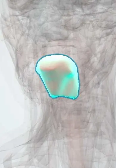
Representative model, for educational purposes only.