Conditions · Ophthalmology and ENT
Thyroglossal duct cyst and branchial cleft cyst !
Neck lumps present from birth that come from embryonic leftovers: a midline cyst near the hyoid or a lateral cyst along the sternocleidomastoid.
Updated 2026-11-01
Structures involved
Tap one to see what it is and open it in 3D.
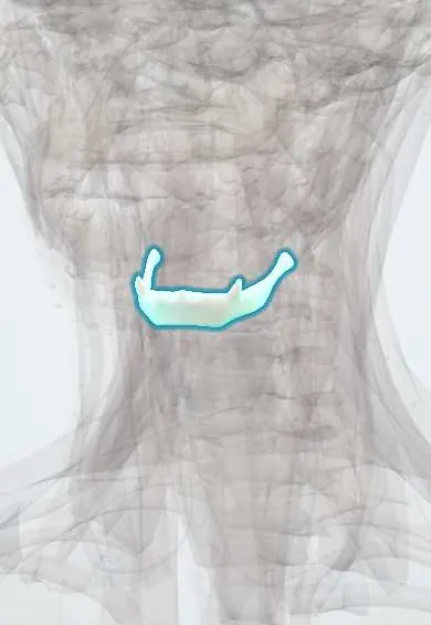
- Hyoid bone
- Thyroid gland
- Sternocleidomastoid
- Tongue
- Thyrohyoid membrane
- Neck lymph nodes
- Internal carotid artery
- External carotid artery
- Palatine tonsils
What it is
The two most common congenital neck masses. A thyroglossal duct cyst is a remnant of the tract along which the thyroid descends in the embryo and appears in the midline. A branchial (pharyngeal) cleft cyst is a remnant of the embryonic pharyngeal apparatus and appears on the side of the neck, usually in front of the sternocleidomastoid.
Who gets it
Thyroglossal duct cysts are the most common congenital neck mass and usually show up in children, often after a respiratory infection. Branchial cleft anomalies come next and often present in older children or young adults. Both are seen worldwide without specific Colombian figures.
Causes
- Thyroglossal duct cyst: persistence of part of the thyroglossal duct between the foramen caecum of the tongue and the thyroid gland
- Branchial cleft cyst: incomplete obliteration of a pharyngeal cleft or pouch, most often the second
- Infection of the cyst often follows an upper respiratory infection
Risk factors
- Congenital, present from birth though often noticed later
- Upper respiratory infections trigger enlargement or infection
Pathophysiology
The thyroid descends from the base of the tongue to the lower neck, passing close to the hyoid bone; if the duct does not involute, retained epithelium secretes fluid and forms a cyst attached to the hyoid, so it moves up when the tongue is protruded or on swallowing. A second branchial cleft remnant can run from the skin in front of the sternocleidomastoid, between the internal and external carotid arteries, to the tonsillar fossa, forming a cyst, sinus or fistula.
Classification
- Thyroglossal duct cyst (midline, at or below the hyoid)
- Branchial cleft anomalies: first (near the ear and parotid), second (most common, anterior border of the sternocleidomastoid), third and fourth (lower neck, may cause recurrent thyroiditis)
- Cyst, sinus or fistula
Symptoms and signs
- Thyroglossal duct cyst: painless, round, midline neck lump near the hyoid that moves up with tongue protrusion and swallowing
- Branchial cleft cyst: smooth, painless lateral neck lump along the anterior border of the sternocleidomastoid
- A small skin pit or opening that drains mucus (sinus or fistula)
- Sudden enlargement, pain, redness and fever when infected
Diagnosis
- Clinical
- Location is the key: midline mass that rises with tongue protrusion suggests a thyroglossal duct cyst; lateral mass along the sternocleidomastoid suggests a branchial cleft cyst.
- Tests
- Thyroid function tests; fine-needle aspiration in adults or atypical lesions to exclude malignancy (papillary thyroid carcinoma can arise in a thyroglossal cyst).
- Imaging
- Neck ultrasound is first line and confirms a normal thyroid in its usual place before excision; CT or MRI for complex, infected or recurrent lesions and to map a fistula tract.
Differential diagnosis
- Reactive or infected cervical lymph node
- Ectopic thyroid or thyroid nodule
- Dermoid cyst (midline, does not move with tongue)
- Lymphatic malformation (cystic hygroma)
- Cystic metastasis of squamous cell or papillary thyroid carcinoma in an adult
Goal of treatment
Remove the lesion completely to prevent recurrent infection and recurrence, after treating any active infection and confirming normal thyroid tissue.
Treatment
- Medical
- An infected cyst is first treated with antibiotics; incision and drainage are avoided where possible because they make later complete excision harder.
- Surgical
- Thyroglossal duct cyst: Sistrunk procedure, removing the cyst, its tract and the central part of the hyoid bone, which greatly lowers recurrence.
- Surgical
- Branchial cleft cyst: complete excision of the cyst and any sinus or fistula tract, protecting nearby nerves and vessels.
- Follow-up
- After surgery: airway, bleeding and drain checks, wound care, and follow-up for recurrence.
Complications
- Recurrent infection and abscess
- Recurrence after incomplete excision
- Draining fistula to the skin
- Rare malignancy in the cyst (papillary thyroid carcinoma in thyroglossal cysts)
- Nerve injury during surgery (facial, hypoglossal, accessory)
Prognosis
Excellent after complete excision; recurrence is low with the Sistrunk procedure and higher after previous infection or simple cyst removal.
Test yourself
Think of the answer, then tap the question to see it.
Which neck mass moves upward when the tongue is protruded?
A thyroglossal duct cyst, because it is attached to the hyoid and the foramen caecum.
Where does a second branchial cleft cyst typically appear?
On the lateral neck, along the anterior border of the sternocleidomastoid.
What operation treats a thyroglossal duct cyst?
The Sistrunk procedure, which includes the central part of the hyoid bone.
Why is a thyroid ultrasound done before excising a midline cyst?
To confirm normal thyroid tissue in its usual place, since the mass could be the only thyroid tissue.
Related conditions
- Thyroid nodule
- Thyroid cancer (papillary)
- Cervical lymphadenopathy
- Deep neck infection and Ludwig angina
- Hypothyroidism
- Head and neck squamous cell carcinoma (oral cavity and oropharynx, HPV)
Educational information, not medical advice. For any symptom, see a health professional.
Sources: Thyroglossal Duct Cyst (Children's Hospital of Philadelphia, 2026); Branchial cleft cyst (MedlinePlus, US National Library of Medicine, 2026).
Spotted a mistake?We are always improving for the community. Tell us what is wrong and our team will review it.Report a mistake
Representative model, for educational purposes only.