Bell palsy !
Bell palsy is sudden weakness of one side of the face, including the forehead, from a facial nerve problem of unknown cause.
Updated 2026-10-29
Structures involved
Tap one to see what it is and open it in 3D.
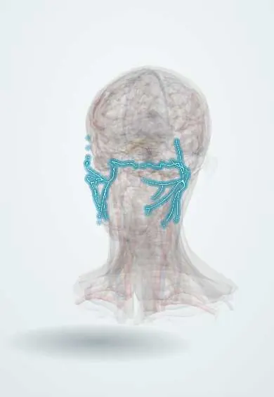
- Facial nerve (VII)
- Facial nerve branches
- Stylomastoid foramen
- Orbicularis oculi
- Orbicularis oris
- Frontalis
- Internal acoustic meatus
- Lacrimal gland
- Temporal bones
In histology
- Peripheral nerve: endoneurium, perineurium, epineurium; myelin
- Nerve injury and regeneration (Wallerian degeneration)
In embryology
- Pharyngeal arches: nerve, muscles, skeleton and artery (the table)
- Second pharyngeal arch (hyoid arch) and the facial nerve
In the clinical exam
In physiology
- Motor cortex and the corticospinal tract
- Upper versus lower motor neuron lesions
- Hearing: middle ear, cochlea and auditory pathway
- Taste and smell
What it is
Sudden weakness or paralysis of one side of the face from an idiopathic lesion of the facial nerve (cranial nerve VII), developing over less than 72 hours. It is a lower motor neuron palsy, so the forehead is affected too.
Who gets it
The commonest cause of acute facial palsy, seen at any age but most often in young and middle-aged adults. More common in pregnancy and in people with diabetes.
Causes
- Idiopathic by definition
- Reactivation of herpes simplex virus type 1 in the geniculate ganglion is the leading hypothesis
Risk factors
- Pregnancy, especially the third trimester and early postpartum
- Diabetes
- Obesity and hypertension
- Recent viral upper respiratory infection
Pathophysiology
Inflammation makes the facial nerve swell inside its narrow bony canal in the temporal bone, especially the labyrinthine segment. The trapped nerve is compressed and ischaemic, causing conduction block or axonal loss. Because all fibres to one half of the face run in this nerve, upper and lower face are both weak, unlike a stroke.
Symptoms and signs
- Weakness of the whole half of the face over hours to a day or two
- Unable to raise the eyebrow or wrinkle the forehead on that side
- Incomplete eye closure; the eye rolls up on trying (Bell phenomenon)
- Drooping mouth corner, drooling, food trapped in the cheek
- Pain behind the ear; altered taste on the front of the tongue
- Sensitivity to loud sounds; dry eye or less tearing
Diagnosis
- Clinical
- A diagnosis of exclusion. Acute onset, whole-face weakness including the forehead and no other neurological signs support it; ear, parotid and skin are examined. Severity is graded, for example with the House–Brackmann scale.
- Tests
- Not routine. Glucose when diabetes is suspected; Lyme serology only where exposure is possible. Electroneurography in complete palsy can help estimate the prognosis.
- Imaging
- Not needed in typical cases. Contrast MRI of the brain and facial nerve path, or CT of the temporal bone, for atypical features, recurrence or no recovery.
Differential diagnosis
- Stroke (forehead spared)
- Ramsay Hunt syndrome (herpes zoster oticus)
- Otitis media, mastoiditis or cholesteatoma
- Parotid tumour or acoustic neuroma
- Lyme disease, Guillain–Barré syndrome, sarcoidosis (often bilateral)
- Temporal bone fracture
Goal of treatment
Improve the chance of full recovery by reducing nerve inflammation early, protect the cornea while the eye cannot close, and support recovery of facial function.
Treatment
- Medical
- Oral corticosteroids started within 72 hours of onset: the treatment with the strongest evidence for improving recovery.
- Medical
- Antivirals may be added to corticosteroids in severe or complete palsy; their benefit is uncertain and guidelines differ. Antivirals alone are not recommended.
- Conservative
- Eye protection: lubricating drops by day, ointment at night, taping the lid closed or a moisture chamber, and protective glasses.
- Rehabilitation
- Facial physiotherapy and exercises may help selected patients; evidence is limited.
- Surgical
- For persistent weakness: eyelid procedures (gold or platinum weight), static or dynamic facial reanimation; routine nerve decompression is not recommended.
Complications
- Exposure keratitis and corneal ulceration
- Incomplete recovery with lasting asymmetry
- Synkinesis (e.g. eye closing when smiling)
- Gustatory tearing («crocodile tears»)
- Facial muscle contracture
- Psychological distress
Prognosis
Most people recover fully, usually starting within 3 weeks and largely within 3 months, especially when the palsy is incomplete. Complete palsy, older age, diabetes and lack of early improvement predict incomplete recovery.
Test yourself
Think of the answer, then tap the question to see it.
How does facial weakness in Bell palsy differ from that of a stroke?
Bell palsy is lower motor neuron and involves the forehead; a stroke usually spares the forehead because its muscles have bilateral cortical input.
Which treatment within 72 hours of onset improves recovery in Bell palsy?
Oral corticosteroids.
Why is eye care essential in Bell palsy?
Incomplete eye closure and less tearing expose the cornea to drying, keratitis and ulceration.
Which syndrome causes facial palsy with vesicles in the ear?
Ramsay Hunt syndrome (herpes zoster oticus).
Where does the facial nerve leave the skull to reach the face?
Through the stylomastoid foramen, then it divides into its branches within the parotid gland.
Related conditions
- Acute otitis media
- Type 2 diabetes mellitus
- Ischaemic stroke
- Parotid gland tumours
- Herpes zoster (shingles)
- Guillain-Barré syndrome
Educational information, not medical advice. For any symptom, see a health professional.
Sources: Bell Palsy (StatPearls (NCBI Bookshelf), 2024).
Spotted a mistake?We are always improving for the community. Tell us what is wrong and our team will review it.Report a mistake
Representative model, for educational purposes only.