Conditions · Hematology and oncology
Hereditary haemorrhagic telangiectasia (Osler–Weber–Rendu) !
An inherited blood vessel disorder that causes repeated nosebleeds, red spots on lips and fingers, and abnormal vessels in the lungs, brain and liver.
Updated 2026-11-01
Structures involved
Tap one to see what it is and open it in 3D.
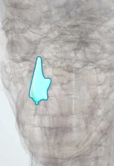
- Nasal septum
- Liver
- Nasal cavity
- Tongue
- Pulmonary arteries
- Pulmonary veins
- Hepatic veins
- Portal vein
- Stomach
- Duodenum
Why it happens
- Mendelian inheritance: autosomal dominant, recessive and X-linked
- Hemorrhage: petechiae, purpura, ecchymosis, hematoma, and what blood loss does
- Blood vessels: artery, vein and capillary layers
What it is
An autosomal dominant disorder of blood vessel development in which arteries connect directly to veins without capillaries. Small connections in the skin and mucosa form telangiectasias that bleed easily; larger arteriovenous malformations (AVMs) occur in the lungs, brain, liver and gut.
Who gets it
It affects roughly 1 in 5,000 to 10,000 people in all ethnic groups, men and women equally. Nosebleeds usually begin in childhood, skin telangiectasias appear by adulthood, and many patients go years without a diagnosis because nosebleeds are so common in the general population. Latin American series describe the same features; in Colombia it is often first recognised in otorhinolaryngology clinics.
Causes
- Pathogenic variants in ENG (HHT type 1) or ACVRL1/ALK1 (HHT type 2)
- SMAD4 variants cause combined juvenile polyposis and HHT
- GDF2 and other rarer genes
- Autosomal dominant inheritance: each child of an affected parent has a 50% chance
Risk factors
- A parent, sibling or child with HHT
- Dry air, nasal trauma and anticoagulant or antiplatelet drugs worsen bleeding
- Pregnancy raises the risk of complications from lung AVMs
Pathophysiology
The affected genes encode parts of the TGF-beta / BMP9 signalling pathway in endothelial cells, which controls how new vessels mature. With half the normal signal, vessels exposed to a second trigger (injury, inflammation) grow into dilated, thin-walled direct artery-to-vein channels. In the nose and gut these rupture with minor trauma. Lung AVMs form a right-to-left shunt that lets clots and bacteria bypass the lung filter, causing stroke and brain abscess, and they lower oxygen saturation. Liver AVMs can cause high-output heart failure, portal hypertension or biliary ischaemia.
Classification
- HHT1 (ENG): more lung and brain AVMs, earlier symptoms
- HHT2 (ACVRL1): more liver AVMs
- Juvenile polyposis–HHT overlap (SMAD4)
- Curaçao clinical criteria: definite with 3 or 4, possible with 2 of recurrent spontaneous epistaxis, mucocutaneous telangiectasias, visceral AVMs and a first-degree relative with HHT
Symptoms and signs
- Recurrent spontaneous nosebleeds, often from childhood and the most common symptom
- Small red or purple spots on the lips, tongue, face, fingertips and nasal mucosa that blanch with pressure
- Tiredness and pallor from iron-deficiency anaemia
- Black stools or gut bleeding, mostly after age 50
- Breathlessness, cyanosis or clubbing from lung AVMs
- Headache or seizures from brain AVMs
Diagnosis
- Clinical
- Based on the Curaçao criteria and a careful family history; examine lips, tongue, palate, fingers and nasal mucosa. Once suspected, patients are screened for visceral AVMs even without symptoms.
- Tests
- Full blood count and ferritin for iron-deficiency anaemia; pulse oximetry; genetic testing for ENG, ACVRL1 and SMAD4 confirms the diagnosis and allows family screening. Endoscopy for gut bleeding, and colonoscopy for polyps in SMAD4 carriers.
- Imaging
- Transthoracic contrast (bubble) echocardiography to screen for lung AVMs, followed by chest CT when positive; brain MRI to screen for cerebral AVMs; Doppler ultrasound or CT of the liver when liver AVMs are suspected.
Differential diagnosis
- Common anterior epistaxis without systemic disease
- Bleeding disorders such as von Willebrand disease
- CREST/limited systemic sclerosis (telangiectasias)
- Spider angiomas of liver disease
- Isolated angiodysplasia of the gut
- Sporadic cerebral or pulmonary AVM
Goal of treatment
Reduce nosebleeds and gut bleeding, correct iron deficiency, and find and treat lung and brain AVMs before they cause stroke, abscess or haemorrhage.
Treatment
- Conservative
- Nasal moisturisers, saline and humidification to protect the mucosa; gentle compression for acute nosebleeds and avoiding nasal packing that tears vessels when possible.
- Medical
- Iron replacement (oral or intravenous) and transfusion for anaemia; antifibrinolytics such as tranexamic acid, and anti-angiogenic therapy (bevacizumab) or other agents for severe bleeding in specialist centres.
- Procedure
- Laser or other ablation of nasal and gut telangiectasias; septodermoplasty or nasal closure for refractory epistaxis.
- Procedure
- Transcatheter embolisation of lung AVMs with a feeding artery large enough to treat; brain AVMs are managed by neurosurgery, embolisation or radiosurgery depending on risk.
- Follow-up
- Antibiotic prophylaxis before dental and other procedures and air filters on intravenous lines when lung AVMs are present; repeat screening and family testing; nursing care includes teaching first aid for nosebleeds and monitoring haemoglobin.
Complications
- Chronic iron-deficiency anaemia and transfusion dependence
- Ischaemic stroke and brain abscess from paradoxical embolism
- Brain haemorrhage
- Hypoxaemia and haemothorax from lung AVMs
- High-output heart failure, portal hypertension and pulmonary hypertension from liver AVMs
- Colorectal cancer risk in SMAD4 juvenile polyposis
Prognosis
Life expectancy is close to normal when visceral AVMs are found and treated early, but quality of life is often limited by bleeding and anaemia. Untreated lung AVMs carry a substantial risk of stroke and brain abscess.
Prevention
- Screening of affected people and their relatives for lung and brain AVMs
- Antibiotic prophylaxis before dental procedures when lung AVMs are present
- Genetic counselling for families
Test yourself
Think of the answer, then tap the question to see it.
What is the inheritance pattern of hereditary haemorrhagic telangiectasia?
Autosomal dominant (ENG, ACVRL1, SMAD4).
What is the most common symptom of HHT?
Recurrent spontaneous nosebleeds (epistaxis).
Why do lung AVMs in HHT cause stroke and brain abscess?
They form a right-to-left shunt that lets clots and bacteria bypass the lung capillaries and reach the brain.
What test screens for pulmonary AVMs in HHT?
Transthoracic contrast (bubble) echocardiography, confirmed by chest CT.
What are the four Curaçao criteria?
Recurrent spontaneous epistaxis, mucocutaneous telangiectasias, visceral AVMs and a first-degree relative with HHT.
Related conditions
- Epistaxis (nosebleed)
- Brain arteriovenous malformation
- Intracerebral haemorrhage
- Brain abscess
- Ischaemic stroke
- Iron deficiency anaemia
- Upper gastrointestinal bleeding
- Heart failure
- Pulmonary hypertension and cor pulmonale
- Portal hypertension
- Colorectal polyps and hereditary polyposis syndromes (FAP, Lynch)
Educational information, not medical advice. For any symptom, see a health professional.
Sources: Hereditary hemorrhagic telangiectasia (MedlinePlus Genetics (U.S. National Library of Medicine), 2020); About Hereditary Hemorrhagic Telangiectasia (HHT) (Centers for Disease Control and Prevention (CDC), 2024).
Spotted a mistake?We are always improving for the community. Tell us what is wrong and our team will review it.Report a mistake
Representative model, for educational purposes only.