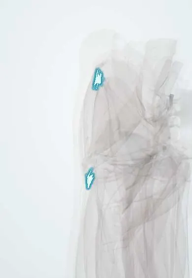
TFL tendon !
The tensor fasciae latae tendon is where this small hip muscle blends into the iliotibial band on the outer thigh.
Official name: Tendon of tensor fasciae latae
Opens in the Durgical app, no sign-up needed.
What it is
The end of the TFL, where it blends into the IT band below the hip.
What it does
Passes the pull of the TFL into the IT band.
Key facts
- Attaches
- The tensor fasciae latae ends between the two layers of the iliotibial tract, about a third of the way down the thigh; the tract continues to Gerdy's tubercle
- Function
- Tenses the iliotibial tract; helps hip flexion, abduction and medial rotation; steadies the extended knee
- Tight in
- Hip extension with adduction (Ober test position)
Relations
- Medial
- Sartorius, diverging from it below the ASIS, with the rectus femoris appearing between them; the lateral femoral cutaneous nerve lies close by
- Deep
- Front parts of the gluteus medius and gluteus minimus; the ascending branch of the lateral circumflex femoral artery
- Behind
- Gluteus maximus, whose fibres join the IT band from behind
- Below
- IT band, continuing down to Gerdy's tubercle
How to find it
With the hip bent and turned inward against resistance, the tensor fasciae latae bulges just below and behind the ASIS; its end merges into the IT band on the upper outer thigh.
Examination
- Ober test: lying on the side, the upper leg is extended and abducted, then allowed to drop; failure to drop toward the table suggests a tight tensor fasciae latae and IT band
On imaging
MRI and ultrasound show the iliotibial band as a thin dark band along the lateral thigh to Gerdy's tubercle. In iliotibial band syndrome the band itself may look normal or thick, with fluid or oedema between it and the lateral femoral epicondyle. Dynamic ultrasound shows the band flicking over the greater trochanter in an external snapping hip. Radiographs are usually normal.
Common problems
- Overuse pain at the front-outer hip, often when it works too hard to make up for a weak gluteus medius.
Clinical notes
- Strips of fascia lata from below it are used as autografts, and the muscle can be raised as a flap on its vascular pedicle
- Iliotibial band syndrome: in runners and cyclists, lateral knee pain where the band crosses the lateral femoral epicondyle, worst with the knee bent about 30°
Anatomical variants
- In about a third of people the muscle reaches below the greater trochanter and has to be split in a lateral approach
Test yourself
Think of the answer, then tap the question to see it.
Where does the tensor fasciae latae end?
Between the two layers of the iliotibial tract, about a third of the way down the thigh; the tract continues to Gerdy's tubercle.
Which interval does the direct anterior hip approach use, and which nerve is at risk?
Between the tensor fasciae latae (superior gluteal nerve) and the sartorius (femoral nerve); the lateral femoral cutaneous nerve is at risk.
What does a positive Ober test suggest?
A tight tensor fasciae latae and IT band: the extended, abducted upper leg fails to drop toward the table.
Educational information, not medical advice. For any symptom, see a health professional.
Its names
- Official Latin (TA2)
- tensor fasciae latae TA2 2602
- Official Spanish (DeCS)
- Tendón del tensor de la fascia lata DeCS D013710
- English (TA2)
- Tendon of tensor fasciae latae
- Also called
- tensor fasciae latae muscle · tensor of fascia lata
Related
Sources: Terminologia Anatomica (TA2); DeCS; Anatomy, Bony Pelvis and Lower Limb: Tensor Fasciae Latae Muscle.
Spotted a mistake?We are always improving for the community. Tell us what is wrong and our team will review it.Report a mistake
Representative model, for educational purposes only.