Nervous system · Head and neck
Cerebellar tonsils !
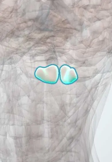
The cerebellar tonsils are two rounded lobes on the underside of the cerebellum, just above the large opening of the skull base, into which they can be pushed.
Official name: Tonsil of cerebellum
Opens in the Durgical app, no sign-up needed.
What it is
Two rounded lobules on the underside of the cerebellum, above the foramen magnum.
What it does
Can be pushed down into the foramen magnum (Chiari malformation, herniation).
Key facts
- Where
- Underside of each cerebellar hemisphere, next to the foramen magnum
- Blood supply
- Posterior inferior cerebellar artery (PICA)
- Function
- Part of the cerebellum; can herniate through the foramen magnum and press on the medulla (Chiari I: more than 5 mm below)
Relations
- In front
- Medulla oblongata, with the posterior inferior cerebellar artery looping between them
- Medial
- The other tonsil, separated by the vallecula, where the inferior vermis and the median aperture of the fourth ventricle lie
- Below
- Rim of the foramen magnum
- Behind
- Cisterna magna and the inner surface of the occipital bone
- Above
- Inferior medullary velum and the uvula of the vermis; the tonsil lies outside the fourth ventricle, below its lower roof
How to find it
On a sagittal MRI the tips of the tonsils are compared with a line joining the front and back edges of the foramen magnum (basion to opisthion); tips more than about 5 mm below it are the usual threshold for Chiari I. At surgery they appear beneath the cisterna magna once the posterior fossa dura is opened.
On imaging
Midline sagittal MRI: the tip of each tonsil is compared with a line from the front to the back edge of the foramen magnum. CSF flow studies show whether flow around them is blocked; a cord syrinx is looked for in the same scan.
Clinical notes
- Symptomatic Chiari I is treated by posterior fossa decompression: removing bone from the back of the foramen magnum and the arch of the atlas, often widening the dura
- A spinal CSF leak (low pressure) makes the brain sag and the tonsils descend, mimicking Chiari I
Anatomical variants
- Tonsils lying a few millimetres below the foramen magnum without symptoms (tonsillar ectopia) are a common chance finding on MRI, especially in children and young adults, and are not treated by themselves
Development
Chiari I is thought to arise mainly from a small, underdeveloped posterior fossa (occipital bone) that leaves too little room for a normal-sized cerebellum, rather than from the tonsils themselves.
Related conditions
Test yourself
Think of the answer, then tap the question to see it.
What is tonsillar herniation (coning), and what are its signs?
Raised pressure pushes the cerebellar tonsils through the foramen magnum onto the medulla: neck stiffness, slowing pulse, rising blood pressure, irregular breathing, then arrest.
Why is imaging done before lumbar puncture when raised pressure or a posterior fossa mass is suspected?
Because of the risk of tonsillar herniation through the foramen magnum.
What is Chiari I malformation?
Low, pointed cerebellar tonsils (more than 5 mm below the foramen magnum) causing cough-induced occipital headache and a cervical syrinx.
Educational information, not medical advice. For any symptom, see a health professional.
Its names
- Official Latin (TA2)
- tonsilla cerebelli TA2 5817
- Official Spanish (DeCS)
- Amígdalas
- English (TA2)
- Tonsil of cerebellum
- Also called
- lobule IX of cerebellar hemisphere · lobule H IX
Related
Sources: Terminologia Anatomica (TA2); Neuroanatomy, Cerebellum; Tratado de anatomía humana (9.ª ed.), tomo II, Libro V, sección 2 (Encéfalo), capítulos I-V: bulbo, protuberancia, cerebelo, cuarto ventrículo, mesencéfalo.
Spotted a mistake?We are always improving for the community. Tell us what is wrong and our team will review it.Report a mistake
Representative model, for educational purposes only.