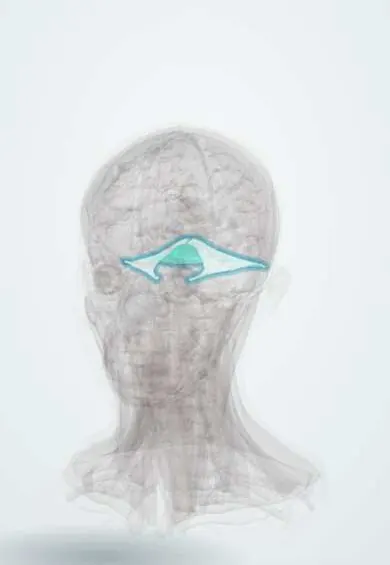
Raised intracranial pressure and brain herniation !
Raised intracranial pressure is high pressure inside the skull from swelling, bleeding, a mass or fluid. It can push the brain out of place (herniation).
Updated 2026-11-01
Structures involved
Tap one to see what it is and open it in 3D.
- Tentorium
- Cerebellar tonsils
- Midbrain
- Lateral ventricles
- Falx cerebri
- Foramen magnum
- Medulla oblongata
- Oculomotor nerve (III)
- Optic nerve
- Third ventricle
- Cerebral aqueduct
- Choroid plexus
- Cingulate gyrus
In embryology
In the clinical exam
- Fundoscopy: the optic disc, papilledema and retinopathy
- Pupils: light reflex and abnormal pupils (Marcus Gunn, Horner, Argyll Robertson)
- Intracranial hypertension syndrome
- Meningeal syndrome
Why it happens
- Edema and effusions: the four mechanisms
- Cerebral blood flow, brain metabolism and intracranial pressure
- CSF and the blood–brain barrier
- Blood–brain barrier, choroid plexus and cerebrospinal fluid
What it is
A sustained rise in the pressure inside the skull, usually above 20–22 mmHg in adults, caused by extra volume of brain tissue, blood or cerebrospinal fluid. If it keeps rising, brain tissue is pushed across the dural folds or through the foramen magnum (herniation), a life-threatening emergency.
Who gets it
A common final pathway in neurocritical care. In Colombia and Latin America, traumatic brain injury from road traffic collisions (especially motorcycles) and violence is a leading cause in young adults; stroke, intracranial haemorrhage, tumours, central nervous system infections and neurocysticercosis with hydrocephalus are other frequent causes.
Causes
- Mass lesions: epidural, subdural or intracerebral haematoma, tumour, abscess
- Cerebral oedema: traumatic brain injury, large ischaemic stroke, hypoxic injury, liver failure, hyponatraemia
- Cerebrospinal fluid build-up: hydrocephalus, including obstruction by neurocysticercosis
- Infection: meningitis and encephalitis
- Increased blood volume: venous sinus thrombosis, hypercapnia, severe hypertension
- Idiopathic intracranial hypertension (a chronic form, mainly in young women with obesity)
Risk factors
- Severe head trauma
- Large stroke or intracranial bleeding
- Brain tumours
- Central nervous system infections
- Hypoventilation with retained carbon dioxide
Pathophysiology
The skull is a fixed box holding brain, blood and cerebrospinal fluid (Monro-Kellie doctrine). At first a new volume is offset by shifting CSF and venous blood out, but once this reserve is used up small additions cause steep rises in pressure. Cerebral perfusion pressure (mean arterial pressure minus intracranial pressure) then falls, causing ischaemia, more oedema and a vicious cycle. Pressure gradients push tissue: the cingulate gyrus under the falx (subfalcine), the medial temporal lobe (uncus) over the tentorium compressing the oculomotor nerve and midbrain (transtentorial), and the cerebellar tonsils into the foramen magnum compressing the medulla (tonsillar).
Classification
- Subfalcine herniation: cingulate gyrus under the falx; can compress the anterior cerebral artery
- Uncal (lateral transtentorial) herniation: ipsilateral dilated pupil from oculomotor nerve compression, then hemiparesis and coma
- Central (downward transtentorial) herniation: small pupils, Cheyne-Stokes breathing, decorticate then decerebrate posturing
- Tonsillar herniation: cerebellar tonsils through the foramen magnum, respiratory arrest
- Upward and external (through a skull defect) herniation
Symptoms and signs
- Headache, worse in the morning, on lying down or with coughing and straining
- Vomiting, often without nausea
- Drowsiness, confusion and a falling Glasgow Coma Scale score
- Blurred or double vision; papilloedema on fundoscopy
- Abducens (sixth) nerve palsy as a false localising sign
- In infants: bulging fontanelle, irritability and enlarging head
Diagnosis
- Clinical
- Suspected from the history (trauma, stroke, tumour, infection), a falling level of consciousness, pupil changes and focal signs. Serial Glasgow Coma Scale, pupil size and reactivity, and vital signs track progression. Fundoscopy may show papilloedema, though it can be absent early.
- Tests
- Invasive ICP monitoring with an intraventricular catheter (which also drains CSF) or an intraparenchymal probe in severe cases; blood gases, sodium and osmolality, glucose, clotting and blood count. Lumbar puncture is avoided when a mass effect is present because it can precipitate herniation.
- Imaging
- Urgent non-contrast head CT shows the cause and signs of mass effect: midline shift, effaced sulci and basal cisterns, compressed ventricles or hydrocephalus. MRI and CT or MR venography are used for tumours, infection and venous thrombosis. Optic nerve sheath ultrasound is a bedside adjunct.
Differential diagnosis
- Postictal state or non-convulsive status epilepticus
- Metabolic or toxic encephalopathy (hypoglycaemia, uraemia, liver failure, drugs)
- Brainstem stroke
- Migraine with severe headache
- Pseudopapilloedema (optic disc drusen)
Goal of treatment
Keep cerebral perfusion pressure adequate, lower intracranial pressure, prevent or reverse herniation and treat the cause (evacuate a haematoma, drain CSF, remove a mass, treat infection).
Treatment
- Acute
- First measures: secure the airway (intubation if the Glasgow Coma Scale is 8 or less), avoid hypoxia and hypotension, head of the bed raised to about 30 degrees with the head midline, and avoid tight neck ties so venous return is not blocked.
- Acute
- Herniation emergency: hyperosmolar therapy (mannitol or hypertonic saline) and brief controlled hyperventilation only as a bridge to definitive treatment, because prolonged hypocapnia causes cerebral ischaemia.
- Medical
- Supportive medical care: sedation and analgesia, normothermia, normoglycaemia and normal sodium, seizure treatment and prevention when indicated; glucocorticoids reduce oedema around tumours and abscesses but are not used in traumatic brain injury.
- Procedure
- External ventricular drain to remove CSF and measure pressure, especially in hydrocephalus.
- Surgical
- Surgery for the cause: evacuation of a haematoma, resection of a mass, or decompressive craniectomy for refractory pressure.
- Follow-up
- Nursing priorities: hourly neurological checks with pupils and Glasgow Coma Scale, watch for the Cushing triad, cluster care and keep the room calm, avoid coughing, straining, hip flexion and excessive suctioning, and keep the drain at the ordered level.
Complications
- Brain herniation with brainstem compression and death
- Global or regional cerebral ischaemia
- Permanent neurological deficits and disability
- Vision loss from long-standing papilloedema
- Brain death
Prognosis
Depends on the cause, how high the pressure rises and how quickly it is controlled. Pressure that stays high despite treatment and signs of herniation carry a poor outlook, while early relief of a surgical cause such as an epidural haematoma can lead to a good recovery.
Prevention
- Helmet use on motorcycles and bicycles and seat belts to prevent head injury
- Control of blood pressure to reduce stroke and haemorrhage
- Early treatment of meningitis and of neurocysticercosis
Test yourself
Think of the answer, then tap the question to see it.
What are the three signs of the Cushing triad?
Hypertension (often with a widened pulse pressure), bradycardia and irregular breathing; it signals impending herniation.
How is cerebral perfusion pressure calculated?
Mean arterial pressure minus intracranial pressure.
Which cranial nerve sign is typical of uncal herniation?
An ipsilateral fixed, dilated pupil from compression of the oculomotor nerve (III).
Why is lumbar puncture avoided when a mass lesion raises intracranial pressure?
Lowering pressure below the lesion can provoke downward herniation through the tentorium or foramen magnum.
Which two nursing measures help lower intracranial pressure?
Head of the bed at about 30 degrees with the neck midline, and avoiding activities that raise pressure such as straining, coughing, hip flexion and prolonged suctioning.
Related conditions
- Traumatic brain injury
- Epidural haematoma
- Subdural haematoma
- Intracerebral haemorrhage
- Subarachnoid haemorrhage
- Hydrocephalus
- Glioblastoma
- Bacterial meningitis
- Ischaemic stroke
- Neurocysticercosis
- Status epilepticus
Educational information, not medical advice. For any symptom, see a health professional.
Sources: Increased intracranial pressure: MedlinePlus Medical Encyclopedia (MedlinePlus, US National Library of Medicine, 2026); Brain herniation: MedlinePlus Medical Encyclopedia (MedlinePlus, US National Library of Medicine, 2026).
Spotted a mistake?We are always improving for the community. Tell us what is wrong and our team will review it.Report a mistake
Representative model, for educational purposes only.