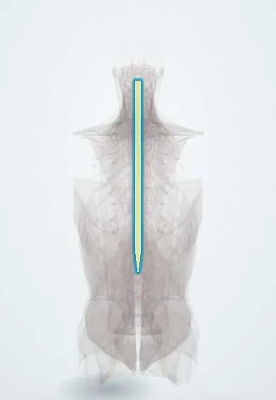
Syringomyelia and Chiari malformation !
In Chiari malformation the lower cerebellum sits in the spinal canal; this can block spinal fluid and form a cavity (syrinx) inside the cord.
Updated 2026-11-01
Structures involved
Tap one to see what it is and open it in 3D.
- Spinal cord
- Cerebellar tonsils
- Foramen magnum
- Fourth ventricle
- Medulla oblongata
- Cerebellum
- Base of the skull
- Atlas (C1)
- Axis (C2)
- Neck nerve roots
Why it happens
What it is
Chiari malformation type I is a downward displacement of the cerebellar tonsils through the foramen magnum. Syringomyelia is a fluid-filled cavity (syrinx) inside the spinal cord, most often in the cervical cord, and Chiari I is its most common cause.
Who gets it
Chiari I is often found incidentally on MRI and many people never have symptoms. Syringomyelia usually becomes symptomatic between 25 and 40 years and is somewhat more frequent in men. Chiari II occurs almost always with myelomeningocele, which remains relevant in Latin America, where neural tube defects are prevented through folic acid fortification and supplementation.
Causes
- Chiari I: small posterior fossa causing tonsillar descent, usually congenital
- Chiari II: descent of the vermis and brainstem, associated with myelomeningocele and hydrocephalus
- Syringomyelia secondary to Chiari malformation (most common)
- Post-traumatic syringomyelia after spinal cord injury
- Syringomyelia from spinal cord tumours or arachnoiditis after infection or haemorrhage
Risk factors
- Neural tube defects (myelomeningocele) for Chiari II
- Prior spinal cord injury or spinal infection
- Connective tissue disorders and craniovertebral junction anomalies
- Acquired tonsillar descent after repeated lumbar CSF drainage (rare)
Pathophysiology
The tonsils crowd the foramen magnum and obstruct the normal pulsatile flow of cerebrospinal fluid between the skull and the spinal canal. Pressure waves drive fluid into the cord, forming a syrinx that usually starts in the central cervical cord. The expanding cavity first interrupts the crossing spinothalamic fibres in the anterior commissure, causing loss of pain and temperature in a cape distribution with preserved touch and proprioception, then damages anterior horn cells and the long tracts.
Classification
- Chiari type I: tonsils at least 5 mm below the foramen magnum
- Chiari type II: vermis, brainstem and fourth ventricle displaced, with myelomeningocele
- Chiari type III: herniation of hindbrain through an occipital or high cervical encephalocele (rare)
- Syringomyelia: communicating, non-communicating (Chiari, trauma, tumour) or atrophic
Symptoms and signs
- Occipital headache brought on by coughing, sneezing or straining (Chiari I)
- Neck pain, dizziness, imbalance and unsteady walking
- Loss of pain and temperature sensation over the shoulders and arms in a cape pattern, with painless burns or cuts
- Weakness and wasting of the hand muscles with reduced arm reflexes
- Spasticity and brisk reflexes in the legs
- Scoliosis, especially in children
- Trouble swallowing, hoarseness or sleep apnoea when the brainstem is involved
Diagnosis
- Clinical
- History of cough headache and examination showing dissociated sensory loss in a cape distribution, lower motor neuron signs in the arms and upper motor neuron signs in the legs, and cerebellar or lower cranial nerve signs.
- Tests
- No blood test is diagnostic. Nerve conduction studies and sleep studies may help in selected cases.
- Imaging
- MRI of the brain and whole spine is the key test: it shows tonsillar descent, a crowded foramen magnum and the syrinx; cine MRI assesses CSF flow. Contrast is used to exclude a tumour. CT myelography is an option when MRI is not possible.
Differential diagnosis
- Cervical spondylotic myelopathy
- Intramedullary spinal cord tumour
- Multiple sclerosis or transverse myelitis
- Amyotrophic lateral sclerosis
- Carpal tunnel syndrome or cervical radiculopathy
- Tonsillar descent secondary to low CSF pressure or raised intracranial pressure
Goal of treatment
Restore normal CSF flow at the foramen magnum, stop progression of the syrinx and preserve neurological function; observation is appropriate for people without symptoms.
Treatment
- Follow-up
- Clinical and MRI follow-up for asymptomatic Chiari I or small stable syrinxes.
- Surgical
- Posterior fossa decompression (suboccipital craniectomy with C1 laminectomy, often with duraplasty) is the main treatment for symptomatic Chiari I with or without syrinx. Shunting of the syrinx or treatment of a tumour or tethered cord in selected cases; hydrocephalus is treated first if present.
- Medical
- Analgesics and neuropathic pain agents for pain control.
- Rehabilitation
- Physiotherapy and occupational therapy; education on protecting insensate skin from burns and injuries.
Complications
- Permanent weakness, wasting and sensory loss in the arms
- Chronic neuropathic pain
- Neuropathic (Charcot) joints in the shoulder or elbow
- Progressive scoliosis
- Brainstem dysfunction: dysphagia, sleep apnoea, vocal cord paralysis
- Surgical complications: CSF leak, pseudomeningocele, meningitis, syrinx recurrence
Prognosis
Untreated symptomatic syringomyelia tends to progress slowly over years. Decompression usually halts progression and improves symptoms in most patients, although established sensory loss and wasting often persist.
Prevention
- Folic acid supplementation before conception and in early pregnancy to prevent myelomeningocele and Chiari II
- Prevention of spinal cord injury
Test yourself
Think of the answer, then tap the question to see it.
What is the classic sensory finding in cervical syringomyelia?
Loss of pain and temperature in a cape distribution with preserved touch and proprioception (dissociated sensory loss).
Why are pain and temperature lost first in syringomyelia?
The central syrinx first damages spinothalamic fibres crossing in the anterior white commissure.
Which headache pattern suggests Chiari I malformation?
Occipital headache triggered by coughing, sneezing or straining.
With which congenital defect is Chiari II almost always associated?
Lumbosacral myelomeningocele, usually with hydrocephalus.
What is the main surgical treatment for symptomatic Chiari I?
Posterior fossa (suboccipital) decompression, often with C1 laminectomy and duraplasty.
Related conditions
- Hydrocephalus
- Spina bifida (myelomeningocele)
- Scoliosis
- Spinal cord injury
- Degenerative cervical myelopathy
- Transverse myelitis
- Multiple sclerosis
- Idiopathic intracranial hypertension
Educational information, not medical advice. For any symptom, see a health professional.
Sources: Syringomyelia: MedlinePlus Medical Encyclopedia (MedlinePlus (U.S. National Library of Medicine), 2025); Chiari Malformation | National Institute of Neurological Disorders and Stroke (National Institute of Neurological Disorders and Stroke (NIH), 2025).
Spotted a mistake?We are always improving for the community. Tell us what is wrong and our team will review it.Report a mistake
Representative model, for educational purposes only.