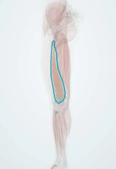
Inclusion body myositis !
A slow muscle disease of people over 50 that weakens the thighs and finger grip, causes falls and swallowing problems, and does not respond to steroids.
Updated 2026-11-01
Structures involved
Tap one to see what it is and open it in 3D.
- Vastus medialis
- FDP
- Vastus lateralis
- Vastus intermedius
- Rectus femoris
- Quadriceps tendon
- Tibialis anterior
- FDS
- FDP tendons
- Pharyngeal constrictors
Why it happens
- Intracellular accumulations: cholesterol, proteins, hyaline, glycogen
- Autophagy
- Creatine kinase (CK) and CK-MB as lab tests
- Protein structure and folding
- Skeletal muscle: sarcomere and contraction
What it is
A chronic inflammatory and degenerative muscle disease and the most common acquired myopathy in people over 50. It produces slowly progressive, often asymmetric weakness of the knee extensors (quadriceps) and the long finger flexors, falls and dysphagia. Unlike dermatomyositis and polymyositis, it does not respond to glucocorticoids or other immunosuppressants.
Who gets it
Estimated prevalence ranges from about 5 to 70 per million, rising sharply after age 50; it is rare before 45. Men are affected about twice as often as women. There are no reliable Colombian data, and diagnosis is often delayed by about five years, frequently after a misdiagnosis of polymyositis.
Causes
- Cause unknown; combines autoimmune attack by cytotoxic T cells with degeneration of muscle fibres
- Abnormal accumulation of misfolded proteins inside fibres (inclusions)
- Possible genetic susceptibility (HLA-DR3 related haplotypes)
- Association with chronic viral infection (HIV, hepatitis C) and with Sjögren disease in some patients
Risk factors
- Age over 50 years
- Male sex
- HIV or hepatitis C infection
- Sjögren disease and other autoimmune diseases
Pathophysiology
CD8 cytotoxic T cells surround and invade non-necrotic muscle fibres that express MHC class I. At the same time the fibres degenerate: they accumulate misfolded proteins (for example p62, TDP-43 and beta-amyloid), develop rimmed vacuoles and show mitochondrial abnormalities. The degenerative component is thought to explain why suppressing inflammation does not stop progression. Autoantibodies against cytosolic 5′-nucleotidase 1A (anti-cN1A) are found in a proportion of patients.
Symptoms and signs
- Slowly progressive weakness over years, often asymmetric
- Weak knee extension: knees buckle, frequent falls, trouble climbing stairs or rising from a chair
- Weak finger flexion: poor grip, trouble turning keys, holding tools or making a fist
- Wasting of the forearm and medial thigh (vastus medialis)
- Foot drop and tripping from ankle dorsiflexor weakness
- Dysphagia in about half of patients, with choking on food
Diagnosis
- Clinical
- Suspect it in anyone over 50 with slowly progressive weakness of the quadriceps and finger flexors (often worse than shoulder abduction), particularly if a diagnosis of polymyositis was made and steroids failed. Diagnosis uses clinical criteria plus biopsy (for example the ENMC criteria).
- Tests
- Creatine kinase is normal or mildly raised (usually less than 15 times normal). Electromyography shows myopathic changes, sometimes mixed with neurogenic-looking potentials. Anti-cN1A antibodies support the diagnosis. Muscle biopsy shows endomysial inflammation with T cells invading fibres, rimmed vacuoles, protein inclusions and COX-negative fibres. Swallowing study (videofluoroscopy) when dysphagia is present.
- Imaging
- Muscle MRI shows a selective pattern of fatty replacement of the quadriceps (especially vastus medialis, with relative sparing of rectus femoris) and the deep finger flexors, and helps choose the biopsy site.
Differential diagnosis
- Polymyositis and other inflammatory myopathies (proximal and symmetric, respond to steroids)
- Amyotrophic lateral sclerosis (fasciculations, upper motor neuron signs)
- Myasthenia gravis (fatigable weakness, ptosis)
- Myotonic dystrophy and other muscular dystrophies
- Hypothyroid or drug-induced (statin) myopathy
- Lumbar radiculopathy or neuropathy causing foot drop
Goal of treatment
There is no cure; care aims to preserve mobility and independence, prevent falls and aspiration, and keep the patient well nourished.
Treatment
- Rehabilitation
- Physiotherapy with regular safe strength and aerobic exercise, ankle-foot orthoses for foot drop, walking aids and home adaptations; occupational therapy with grip aids.
- Rehabilitation
- Speech and swallowing therapy: posture and swallowing manoeuvres, texture-modified diet and nutritional support.
- Procedure
- Procedures for severe dysphagia: dilation or myotomy of the cricopharyngeal muscle; gastrostomy feeding if aspiration persists.
- Medical
- Glucocorticoids and immunosuppressants are generally ineffective and not recommended routinely; intravenous immunoglobulin may give brief benefit for dysphagia in some patients. Treat associated infection or autoimmune disease.
- Follow-up
- Monitoring: fall risk, swallowing screening before meals, weight, respiratory function and signs of aspiration pneumonia; nursing teaches aspiration precautions and safe transfers.
Complications
- Falls with fractures and head injury
- Aspiration pneumonia and malnutrition
- Loss of independent walking (cane, then wheelchair over 10 to 15 years)
- Respiratory muscle weakness late in the disease
- Side effects of unnecessary immunosuppression
Prognosis
Slowly progressive over many years; most patients eventually need a walking aid and many a wheelchair after one to two decades. Life expectancy is close to normal, and the main threats to life are aspiration pneumonia, malnutrition and falls.
Test yourself
Think of the answer, then tap the question to see it.
Which two muscle groups are characteristically weak in inclusion body myositis?
The knee extensors (quadriceps) and the long finger flexors.
What clinical clue suggests IBM in a patient diagnosed with polymyositis?
Failure to respond to glucocorticoids, especially in a man over 50 with asymmetric weakness.
Which biopsy features distinguish IBM?
Rimmed vacuoles and protein inclusions together with endomysial T cells invading non-necrotic fibres.
What is a key nursing priority in IBM?
Preventing falls and aspiration: swallowing screening, texture-modified diet, safe mobility aids.
Related conditions
- Inflammatory myopathies (dermatomyositis, polymyositis)
- Amyotrophic lateral sclerosis
- Myasthenia gravis
- Oropharyngeal dysphagia in older adults
- Aspiration pneumonia and pneumonitis
- Sjögren disease
- HIV infection and AIDS
- Viral hepatitis
- Hypothyroidism
- Peripheral neuropathy (diabetic and other)
- Sarcopenia
- Frailty syndrome
Educational information, not medical advice. For any symptom, see a health professional.
Sources: Inclusion Body Myositis | National Institute of Neurological Disorders and Stroke (National Institute of Neurological Disorders and Stroke (NINDS), 2026); Inclusion Body Myositis - The Myositis Association (The Myositis Association, 2026).
Spotted a mistake?We are always improving for the community. Tell us what is wrong and our team will review it.Report a mistake
Representative model, for educational purposes only.