
Bicuspid aortic valve
About 1 in 100 people are born with an aortic valve of two leaflets instead of three. It wears out decades early and often comes with a widened aorta.
What it is
A congenital aortic valve with two functional leaflets, usually because two of the three cusps are fused along a ridge (raphe). It is the most common congenital heart valve lesion and is often linked to dilatation of the aortic root or ascending aorta (bicuspid aortopathy).
Who gets it
Present in about 1–2% of people, two to three times more often in males. It runs in families and is common in Turner syndrome and in people with coarctation of the aorta. It is the classic cause of aortic stenosis in adults under about 70, and a frequent reason for valve surgery in younger adults.
Causes
- Congenital: the aortic cusps form abnormally early in fetal life
- Familial clustering, with variants in genes such as NOTCH1 and GATA4/GATA6 in some families
- Associated syndromes and lesions: Turner syndrome, coarctation of the aorta and other left-sided obstructive lesions
Risk factors
- Male sex (more stenosis and more root involvement)
- Hypertension
- Smoking and other atherosclerotic risk factors (faster calcification)
- Family history of aortic dissection
- Root phenotype of aortopathy
- Pregnancy with a dilated aorta
Pathophysiology
Two cusps instead of three → the valve opens less widely and its leaflets bend more with each beat → turbulent, off-centre (eccentric) flow and leaflet stress → early fibrosis and calcification (stenosis, typically from the fourth to sixth decade, a decade or two before a three-cusp valve) or incomplete closure (regurgitation, more often in younger patients) → abnormal flow on the aortic wall plus an inherited weakness of the media → progressive dilatation of the aortic root or ascending aorta → aneurysm, with a higher risk of dissection than in the general population, although the absolute risk stays low. The aortopathy can keep progressing after the valve is replaced.
Classification
- By fusion pattern: right–left coronary cusp fusion (most common; dilatation tends to involve the root) and right–non-coronary fusion (dilatation more of the ascending aorta and arch); rarely, a true two-cusp valve without a raphe
- By valve lesion: normal function, stenosis, regurgitation, or both
- By aorta: no dilatation, ascending phenotype, root phenotype (less common, more often in young men, with a lower threshold for surgery)
Symptoms and signs
- Often none for decades; found because of a murmur or an echocardiogram
- An ejection click and a systolic murmur at the upper sternal border
- Exertional breathlessness, angina or syncope once stenosis is severe
- Breathlessness and an early diastolic murmur with regurgitation
- Sudden tearing chest or back pain if the aorta dissects
Diagnosis
- Clinical
- Suspected from an ejection click followed by a systolic murmur, often in a young adult, or in a person with coarctation or Turner syndrome.
- Tests
- ECG for left ventricular hypertrophy; genetic testing only in selected families or syndromes.
- Imaging
- Transthoracic echocardiography shows the two-leaflet valve with an oval 'fish-mouth' opening in systole, measures stenosis and regurgitation, and measures the aortic root and ascending aorta. Cardiac CT or MRI measures the whole thoracic aorta when echo cannot see it well and is used for follow-up; transoesophageal echocardiography when images are poor.
Differential diagnosis
- Degenerative (calcific) stenosis of a three-cusp aortic valve
- Rheumatic aortic valve disease
- Subvalvular or supravalvular aortic stenosis
- Hypertrophic cardiomyopathy with outflow obstruction
- Unicuspid aortic valve
Aortic stenosis: what kind of valve is behind it?
| Cause | Typical age at symptoms | Valve on echo | Clues |
|---|---|---|---|
| Bicuspid aortic valve | 40s to 60s | Two leaflets, fish-mouth opening, often a raphe | Ejection click; dilated ascending aorta; family history |
| Degenerative stenosis of a three-cusp valve | 70s and 80s | Three thickened, calcified cusps | Atherosclerotic risk factors; no aortopathy |
| Rheumatic aortic disease | Young and middle-aged adults where rheumatic fever is common | Fused commissures, often with regurgitation | The mitral valve is almost always affected too |
| Hypertrophic cardiomyopathy | Any age, often young | Normal valve; obstruction below it | Murmur louder on standing or with Valsalva; no ejection click |
Goal of treatment
Follow the valve and the aorta for life, replace or repair the valve when severe disease causes symptoms or harms the ventricle, and replace the aorta before it dissects.
Treatment
- Follow-up
- Lifelong follow-up with echocardiography, and CT or MRI of the aorta when needed, at intervals set by the guideline according to valve function and aortic size.
- Medical
- Blood pressure control and risk-factor control; no drug has been shown to stop valve calcification.
- Surgical
- Aortic valve replacement (surgical, or transcatheter in selected patients) or valve repair for severe stenosis or regurgitation, using the same criteria as for other causes.
- Surgical
- Replacement of the ascending aorta or root when the diameter reaches the threshold set by the guideline; the threshold is lower for the root phenotype, with a family history of dissection or rapid growth, and when valve surgery is being done anyway.
- Lifestyle
- Dental hygiene to lower endocarditis risk; advice on heavy straining and isometric exercise when the aorta is dilated; assessment before pregnancy.
Complications
- Aortic stenosis
- Aortic regurgitation
- Ascending aortic or root aneurysm
- Aortic dissection
- Infective endocarditis
- Left ventricular hypertrophy and heart failure
Common traps
- Once the valve is replaced, the aorta is fixed too: the aortopathy is part of the disease, not only a result of the jet, so the ascending aorta can keep enlarging and still needs follow-up
- Aortic stenosis in a 45-year-old is degenerative: at that age a bicuspid valve is the classic cause; degenerative stenosis of a three-cusp valve presents in the 70s and 80s
- Only the patient needs an echocardiogram: it runs in families, so first-degree relatives are screened for both a bicuspid valve and a dilated aorta
- Once the coarctation is repaired, the heart is done: many people with coarctation also have a bicuspid valve and aortopathy, which need their own lifelong follow-up
Prognosis
Most people live normal lives, but many need valve surgery in adulthood. Dissection is uncommon in absolute terms yet more frequent than in the general population, which is why the aorta is measured regularly.
Prevention
- Echocardiographic screening of first-degree relatives (parents, siblings, children) for both a bicuspid valve and a dilated aorta
- Echocardiography in everyone with coarctation of the aorta or Turner syndrome
- Blood pressure control and not smoking
- Good dental hygiene
Test yourself
Think of the answer, then tap the question to see it.
What is the most common congenital heart valve lesion, and how common is it?
A bicuspid aortic valve, present in about 1–2% of people, more often in males.
Which two problems are followed for life in a person with a bicuspid aortic valve?
The valve (stenosis, regurgitation, endocarditis) and the aorta (dilatation of the root or ascending aorta, with a risk of dissection).
Which conditions should prompt you to look for a bicuspid aortic valve?
Coarctation of the aorta, Turner syndrome, a first-degree relative with a bicuspid valve or thoracic aortic disease, and aortic stenosis in a young or middle-aged adult.
A 22-year-old man has an ejection click and a soft systolic murmur. Echo shows a bicuspid aortic valve with mild stenosis and an ascending aorta at the upper limit of normal. What next?
Explain the diagnosis, plan regular echo follow-up of valve and aorta (CT or MRI if needed), control blood pressure, advise dental hygiene, and offer echocardiography to his parents, siblings and children.
Why it happens
Connections
Educational information, not medical advice. For any symptom, see a health professional.
Sources: Bicuspid Aortic Valve (Archived) (StatPearls (NCBI Bookshelf), 2023); Aortopathy in Bicuspid Aortic Valve: Pathophysiology, Risk Stratification and Surgical Decision-Making—A Narrative Review (Journal of Clinical Medicine (Europe PMC), 2026); Commentary on the 2024 European Society of Cardiology Guidelines on Peripheral Arterial and Aortic Diseases (European Cardiology Review, 2024).
Spotted a mistake?We are always improving for the community. Tell us what is wrong and our team will review it.Report a mistake
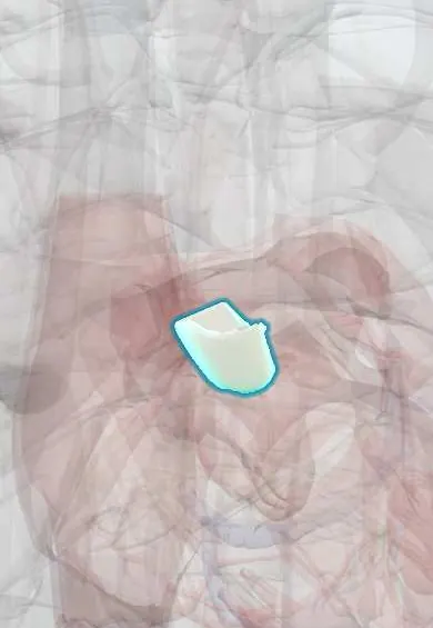
Representative model, for educational purposes only.