Hypertrophic cardiomyopathy !
Hypertrophic cardiomyopathy is an inherited thickening of the heart muscle. It can block blood flow and cause sudden death in young people.
Updated 2026-11-01
Structures involved
Tap one to see what it is and open it in 3D.
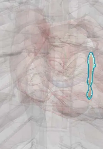
- Interventricular septum
- Left ventricle
- Mitral anterior leaflet
- Left atrium
- Left ventricle papillary muscles
- Mitral posterior leaflet
In the clinical exam
- Precordial inspection and palpation: apex beat, heaves and thrills
- Murmurs: timing, grade, location and radiation
- Valve murmurs: the table to know
- Dynamic auscultation: Valsalva, squatting, standing and handgrip
- Abnormal pulses: paradoxus, alternans, parvus et tardus, bounding
Why it happens
- Mendelian inheritance: autosomal dominant, recessive and X-linked
- Hypertrophy: physiologic and pathologic
- The autopsy: clinical and medicolegal
- The ventricular pressure–volume loop
- Murmurs and the hemodynamics of valve disease
- Cardiac muscle: intercalated discs
What it is
A genetic heart muscle disease with left ventricular hypertrophy, usually asymmetric and involving the septum, that is not explained by abnormal loading such as hypertension or aortic stenosis. Many patients have dynamic obstruction of the left ventricular outflow tract.
Who gets it
One of the most common inherited heart diseases, affecting roughly 1 in 500 people worldwide. It is a well-known cause of sudden cardiac death in young people and athletes; many carriers are unaware of it until family screening or an event.
Causes
- Mutations in sarcomere protein genes, most often beta-myosin heavy chain (MYH7) and myosin-binding protein C (MYBPC3)
- Autosomal dominant inheritance with variable penetrance
- Phenocopies to exclude: Fabry disease, amyloidosis, glycogen storage disorders, athlete's heart
Risk factors
- First-degree relative with hypertrophic cardiomyopathy
- Family history of sudden death at a young age
Pathophysiology
Disorganised, hypertrophied myocytes and fibrosis make the ventricle stiff, causing diastolic dysfunction. In obstructive forms the thick septum and systolic anterior motion (SAM) of the anterior mitral leaflet narrow the outflow tract during systole, which also causes mitral regurgitation. Obstruction worsens when the ventricle is smaller or more forceful (dehydration, Valsalva, standing, vasodilators, inotropes). Myocardial disarray and scar favour ventricular arrhythmias.
Classification
- Obstructive (outflow gradient at rest or provoked) or non-obstructive
- Asymmetric septal, apical or concentric patterns
- End-stage (burnt-out) form with reduced ejection fraction
Symptoms and signs
- Often none; found by family screening or an abnormal ECG
- Exertional dyspnoea and chest pain
- Palpitations
- Dizziness or syncope, especially during or just after exercise
- Harsh systolic murmur at the left sternal border that increases with Valsalva or standing and decreases with squatting or handgrip
Diagnosis
- Clinical
- Family history, symptoms on exertion and a dynamic systolic murmur; manoeuvres that reduce preload (Valsalva, standing) make it louder, unlike aortic stenosis.
- Tests
- ECG: left ventricular hypertrophy, deep narrow Q waves in lateral and inferior leads, deep T-wave inversion in the apical variant. Holter for ventricular arrhythmias; exercise test for symptoms and blood pressure response; genetic testing of the patient and cascade screening of relatives.
- Imaging
- Echocardiography shows wall thickness of 15 mm or more (lower with family history), SAM and the outflow gradient; cardiac MRI defines hypertrophy and fibrosis (late gadolinium enhancement) and helps stratify risk.
Differential diagnosis
- Athlete's heart
- Hypertensive heart disease
- Aortic stenosis
- Cardiac amyloidosis and Fabry disease
- Subaortic membrane
Goal of treatment
Relieve symptoms by lowering the outflow obstruction and improving filling, prevent sudden death in those at high risk, and screen relatives.
Treatment
- Medical
- Negative inotropes and rate slowers: beta-blockers first line, nondihydropyridine calcium-channel blockers (verapamil) or disopyramide as alternatives; cardiac myosin inhibitors (for example mavacamten) in selected obstructive cases.
- Lifestyle
- Avoiding dehydration and agents that worsen obstruction (vasodilators, nitrates, high-dose diuretics, positive inotropes); individualised advice on competitive sport.
- Procedure
- Implantable cardioverter-defibrillator for survivors of cardiac arrest or sustained VT and for those at high estimated risk of sudden death.
- Surgical
- Septal reduction for refractory obstructive symptoms: surgical septal myectomy, or alcohol septal ablation by catheter in selected patients.
- Medical
- Anticoagulation when atrial fibrillation develops, regardless of usual risk scores.
- Follow-up
- Periodic ECG, Holter and echocardiography, risk reassessment, and clinical and echocardiographic screening of first-degree relatives.
Complications
- Sudden cardiac death from ventricular arrhythmias
- Atrial fibrillation and stroke
- Heart failure, including the end-stage dilated form
- Infective endocarditis on the mitral valve
- Mitral regurgitation from systolic anterior motion
Prognosis
Many patients live a normal life span with treatment. Risk stratification, implantable defibrillators and septal reduction have lowered mortality; outcomes are worse with high-risk features or end-stage disease.
Prevention
- Cascade genetic and echocardiographic screening of relatives
- Pre-participation cardiovascular screening of young athletes
Test yourself
Think of the answer, then tap the question to see it.
What is the inheritance pattern of hypertrophic cardiomyopathy?
Autosomal dominant, usually from mutations in sarcomere genes such as MYH7 and MYBPC3.
How does the HCM murmur change with Valsalva and with squatting?
It increases with Valsalva or standing (less preload) and decreases with squatting or handgrip.
What causes outflow tract obstruction in obstructive HCM?
Septal hypertrophy plus systolic anterior motion of the anterior mitral leaflet toward the septum.
What is first-line drug therapy for symptomatic obstructive HCM?
Beta-blockers; verapamil or disopyramide are alternatives, and vasodilators and dehydration are avoided.
Which device prevents sudden death in high-risk HCM?
An implantable cardioverter-defibrillator.
Related conditions
- Cardiac arrest (VF, pulseless VT, asystole, PEA)
- Ventricular tachycardia
- Atrial fibrillation
- Heart failure
- Aortic Valve Stenosis
- Hypertensive heart disease
- Mitral regurgitation
- Amyloidosis
- Dilated cardiomyopathy
Educational information, not medical advice. For any symptom, see a health professional.
Sources: Hypertrophic cardiomyopathy: MedlinePlus Medical Encyclopedia (MedlinePlus, US National Library of Medicine, 2026).
Spotted a mistake?We are always improving for the community. Tell us what is wrong and our team will review it.Report a mistake
Representative model, for educational purposes only.