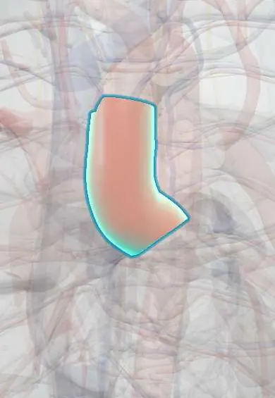
Ascending aorta !
The ascending aorta is the first part of the body's main artery, rising from the left ventricle; the coronary arteries branch from its root.
Opens in the Durgical app, no sign-up needed.
What it is
The first part of the aorta, rising from the left ventricle behind the sternum. The coronary arteries start at its root.
What it does
Carries all the blood the left ventricle pumps out toward the body.
Key facts
- Origin
- Aortic valve of left ventricle, behind left sternum at 3rd intercostal space; inside pericardium, about 5 cm long
- Branches
- Right and left coronary arteries (from aortic sinuses)
- Supplies
- Heart (via coronaries); continues as aortic arch at level of sternal angle
Relations
- In front
- Start of the pulmonary trunk and the right ventricular outflow tract, the right auricle; further forward the pericardium, the thymic remnant and the sternum
- Behind
- Transverse pericardial sinus, then the left atrium, the right pulmonary artery and the right main bronchus
- Lateral
- On the right, the superior vena cava and right atrium; on the left, the pulmonary trunk and the left auricle
- Above
- Becomes the aortic arch at the sternal angle plane (T4–T5), behind the right 2nd sternocostal joint
How to find it
Not palpable. It projects behind the left half of the sternum at the 3rd intercostal space and rises toward the right 2nd costal cartilage; aortic valve sounds and murmurs carry along it to the right 2nd intercostal space at the sternal edge.
On imaging
Contrast CT angiography measures it at the sinuses, the sinotubular junction and mid-ascending level, and shows a dissection flap between true and false lumens; transesophageal echo shows the root and valve. On a frontal chest X-ray a dilated ascending aorta bulges the right upper mediastinal border.
Common problems
- Aneurysm: the wall weakens and widens, usually silently; it can tear (aortic dissection), an emergency with sudden tearing chest pain.
How it is diagnosed
Echocardiogram, CT or MRI.
Treatment
- Blood-pressure control and monitoring; surgical replacement with a graft when it gets too wide.
Clinical notes
- Aneurysm of the root and ascending aorta is linked to Marfan syndrome, bicuspid aortic valve and other connective tissue disorders; a widened root can make the aortic valve leak
- A type A dissection can rupture into the pericardium (tamponade), shear off a coronary ostium (myocardial infarction) or detach the valve (acute aortic regurgitation)
- Site of arterial cannulation and cross-clamping for cardiopulmonary bypass; the proximal ends of vein grafts are sewn onto it in coronary bypass surgery
- Syphilitic aortitis classically affected the ascending aorta, giving a wrinkled tree-bark intima and aneurysm
Development
Forms from the truncus arteriosus, divided from the pulmonary trunk by the spiral aorticopulmonary septum (neural crest cells); failure of the septum leaves a persistent truncus arteriosus.
Related conditions
- Hypertensive emergency and urgency
- Transposition of the great arteries
- Hypoplastic left heart syndrome
- Truncus arteriosus, TAPVR and tricuspid atresia
- Marfan syndrome
- Williams syndrome
- Thoracic aortic aneurysm and aortitis
- Aortic regurgitation
- Tetralogy of Fallot
Test yourself
Think of the answer, then tap the question to see it.
Which arteries arise from the aortic sinuses of the ascending aorta?
The right and left coronary arteries, from the right and left aortic sinuses; the non-coronary sinus gives no artery.
What Stanford type is an aortic dissection that involves the ascending aorta, and how is it managed?
Stanford type A, a surgical emergency.
Which three complications can a type A dissection of the ascending aorta cause near the heart?
Rupture into the pericardium (tamponade), shearing off a coronary ostium (myocardial infarction) and detaching the aortic valve (acute aortic regurgitation).
Which conditions are linked to aneurysm of the aortic root and ascending aorta?
Marfan syndrome, bicuspid aortic valve and other connective tissue disorders; a widened root can make the aortic valve leak.
Educational information, not medical advice. For any symptom, see a health professional.
Its names
- Official Latin (TA2)
- aorta ascendens TA2 4176
- Official Spanish (DeCS)
- Aorta ascendente DeCS D001011
- English (TA2)
- Ascending aorta
Related
Sources: Terminologia Anatomica (TA2); DeCS; Anatomy, Thorax, Heart Great Vessels; Anatomy, Thorax, Mediastinum Superior and Great Vessels.
Spotted a mistake?We are always improving for the community. Tell us what is wrong and our team will review it.Report a mistake
Representative model, for educational purposes only.