Conditions · Surgery and trauma
Thoracic aortic aneurysm and aortitis !
A permanent widening of the aorta in the chest. It is usually silent until it is large, when it can tear or burst; inflamed aorta (aortitis) is one cause.
Updated 2026-11-01
Structures involved
Tap one to see what it is and open it in 3D.
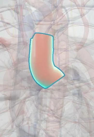
- Ascending aorta
- Aortic arch
- Thoracic aorta
- Aortic right coronary cusp
- Aortic left coronary cusp
- Aortic non-coronary cusp
- Valve rings (fibrous skeleton)
- Brachiocephalic trunk
- Recurrent laryngeal nerves
- Esophagus
- Trachea
- Superior vena cava
- Left ventricle
Why it happens
What it is
A localised permanent dilatation of the thoracic aorta to at least about 1.5 times its expected diameter. It may involve the aortic root and ascending aorta, the arch, or the descending thoracic aorta; thoracoabdominal aneurysms extend below the diaphragm. Aortitis is inflammation of the aortic wall (from large-vessel vasculitis or infection) that can weaken it and lead to aneurysm.
Who gets it
Less common than abdominal aortic aneurysm, with a similar frequency in men and women; incidence rises with age and is often found by chance on imaging. Ascending aneurysms in young people suggest a genetic connective tissue disorder or a bicuspid aortic valve. In Latin America, Takayasu arteritis in young women and syphilitic aortitis are classic causes to remember.
Causes
- Degenerative medial disease (cystic medial degeneration) associated with hypertension and ageing
- Atherosclerosis, mostly in the descending aorta
- Heritable syndromes: Marfan, Loeys-Dietz, vascular Ehlers-Danlos, Turner syndrome, familial thoracic aortic aneurysm
- Bicuspid aortic valve (aortopathy of the ascending aorta)
- Non-infectious aortitis: giant cell arteritis, Takayasu arteritis, other inflammatory diseases
- Infectious aortitis: tertiary syphilis (ascending aorta), bacterial (mycotic) aneurysms, tuberculosis
- Chronic aortic dissection and prior trauma
Risk factors
- Hypertension
- Smoking
- Older age
- Family history of aortic aneurysm or dissection
- Bicuspid aortic valve or coarctation
- Connective tissue disorders
- Stimulant use (cocaine) and heavy weight lifting as triggers of dissection
Pathophysiology
The aortic media loses smooth muscle cells, elastic fibres and collagen organisation, so the wall weakens and gradually dilates. By the law of Laplace, wall tension rises with radius, so dilatation feeds further growth (about a few millimetres per year). In aortitis, granulomatous or lymphoplasmacytic inflammation destroys the media; in syphilis, inflammation of the vasa vasorum causes ischaemic medial damage and a characteristic 'tree-bark' intima. Root dilatation stretches the valve ring and causes aortic regurgitation. A large aneurysm can compress nearby structures and, above all, dissect or rupture.
Classification
- By location: aortic root and ascending, arch, descending thoracic, thoracoabdominal (Crawford types)
- By shape: fusiform (diffuse) or saccular (localised outpouching, more often infective or traumatic)
- By cause: degenerative, genetic, inflammatory (aortitis), infectious (mycotic, syphilitic), post-dissection
- Aortitis: infectious versus non-infectious (large-vessel vasculitis)
Symptoms and signs
- Usually no symptoms; found by chance on chest X-ray, echocardiogram or CT
- Chest or back pain from pressure or expansion
- Hoarseness (stretching of the left recurrent laryngeal nerve)
- Dysphagia, cough, wheeze or stridor from oesophageal or airway compression
- Facial and arm swelling (superior vena cava compression)
- Diastolic murmur and heart failure from aortic regurgitation
- In aortitis: fever, weight loss, raised inflammatory markers, arm claudication and unequal arm pulses or blood pressures
Diagnosis
- Clinical
- Most are found incidentally. Screen first-degree relatives of patients with heritable thoracic aortic disease and people with a bicuspid valve. Look for signs of genetic syndromes (tall stature, lens dislocation, skin fragility) and of vasculitis.
- Tests
- Syphilis serology when the ascending aorta or root is dilated; blood cultures if infection is suspected; ESR and C-reactive protein for aortitis; genetic testing in young patients or with a family history.
- Imaging
- CT angiography or MR angiography measures diameter and extent and plans repair. Transthoracic echocardiography assesses the root, ascending aorta and the aortic valve; transoesophageal echocardiography in selected cases. Chest X-ray may show a widened mediastinum but can be normal. PET-CT can show active aortitis.
Differential diagnosis
- Acute aortic dissection or intramural haematoma
- Acute coronary syndrome
- Mediastinal mass (lymphoma, thymoma, goitre)
- Pulmonary embolism
- Aortic valve disease without aneurysm
- Tortuous but non-dilated (unfolded) aorta
Goal of treatment
Slow growth by controlling blood pressure and risk factors, treat any underlying aortitis, and repair the aorta electively before it reaches the size at which dissection or rupture becomes likely.
Treatment
- Lifestyle
- Stopping smoking, avoiding stimulants and very heavy isometric exertion; blood pressure and lipid control.
- Medical
- Antihypertensive therapy, usually beta-blockers and, in Marfan syndrome, angiotensin receptor blockers. Glucocorticoids and other immunosuppressants for large-vessel vasculitis; penicillin for syphilis; prolonged antibiotics plus surgery for infectious aneurysms.
- Surgical
- Elective open repair of the root or ascending aorta, often with aortic valve repair or replacement, when the diameter reaches about 5.5 cm (lower thresholds in Marfan, Loeys-Dietz, bicuspid valve, rapid growth or symptoms).
- Procedure
- Thoracic endovascular aortic repair (stent graft) for suitable descending thoracic aneurysms, usually from about 5.5–6 cm; hybrid procedures for the arch.
- Follow-up
- Imaging surveillance at intervals set by size and growth; nursing priorities include blood pressure control, education on warning symptoms of dissection, and postoperative neurological checks (spinal cord ischaemia after descending repair).
Complications
- Aortic dissection
- Rupture with massive haemorrhage
- Aortic regurgitation and heart failure
- Compression of airway, oesophagus, recurrent laryngeal nerve or superior vena cava
- Thromboembolism
- After repair: stroke, spinal cord ischaemia with paraplegia, kidney injury
Prognosis
Thoracic aneurysms grow slowly, but the risk of rupture or dissection rises steeply once the diameter passes about 6 cm. Rupture is usually fatal. Elective repair in experienced centres carries a much lower risk than emergency surgery.
Prevention
- Blood pressure control and not smoking
- Screening relatives of patients with heritable thoracic aortic disease
- Echocardiographic follow-up of people with bicuspid aortic valve or Marfan syndrome
- Early treatment of syphilis
Test yourself
Think of the answer, then tap the question to see it.
What is the most common type of pathology underlying a thoracic aortic aneurysm of the ascending aorta?
Degenerative medial disease (cystic medial degeneration), often with hypertension, or a genetic disorder such as Marfan syndrome.
Why can a thoracic aortic aneurysm cause hoarseness?
An arch aneurysm stretches or compresses the left recurrent laryngeal nerve, which loops under the aortic arch.
Which valve lesion is associated with aortic root and ascending aortic dilatation?
Aortic regurgitation from stretching of the valve ring; a bicuspid aortic valve also predisposes to ascending aortopathy.
Which infection classically causes aneurysm of the ascending aorta with 'tree-bark' intima?
Tertiary syphilis, through obliterative inflammation of the vasa vasorum.
Which large-vessel vasculitis typically affects young women and causes unequal arm pulses?
Takayasu arteritis.
Related conditions
- Aortic dissection
- Abdominal aortic aneurysm
- Aortic regurgitation
- Marfan syndrome
- Ehlers–Danlos syndrome
- Turner syndrome
- Syphilis
- Giant cell arteritis
- Takayasu arteritis
- Arterial hypertension
- Aortic Valve Stenosis
- Coarctation of the aorta
Educational information, not medical advice. For any symptom, see a health professional.
Sources: Thoracic Aortic Aneurysms - Cardiology - Merck Manual Professional Edition (Merck Manual Professional Edition, 2025); Thoracic aortic aneurysm: MedlinePlus Medical Encyclopedia (MedlinePlus (U.S. National Library of Medicine), 2024); 2022 ACC/AHA Guideline for the Diagnosis and Management of Aortic Disease (Circulation (American College of Cardiology / American Heart Association), 2022).
Spotted a mistake?We are always improving for the community. Tell us what is wrong and our team will review it.Report a mistake
Representative model, for educational purposes only.