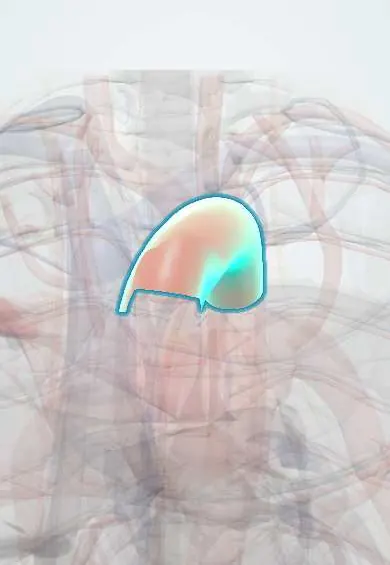
Coarctation of the aorta !
Coarctation of the aorta is a narrowing of the aorta, present from birth, that raises blood pressure in the arms and reduces flow to the legs.
Updated 2026-10-29
Structures involved
Tap one to see what it is and open it in 3D.
- Aortic arch
- Thoracic aorta
- Ligamentum arteriosum
- Subclavian artery
- Posterior intercostal arteries
- Internal thoracic artery
- Left ventricle
- Ribs (back part)
- Ascending aorta
In embryology
In the clinical exam
In physiology
Why it happens
- Sex chromosome disorders: Klinefelter and Turner
- Hypertrophy: physiologic and pathologic
- Systolic, diastolic, mean and pulse pressure
- Fetal circulation and the transition at birth
What it is
A narrowing of the aorta, usually at the isthmus just beyond the left subclavian artery near the ligamentum arteriosum. It raises blood pressure in the arms and reduces flow to the lower body.
Who gets it
More frequent in males and strongly associated with Turner syndrome and a bicuspid aortic valve. It may present as shock in a newborn when the duct closes, or as high blood pressure in a child or young adult.
Causes
- Abnormal development of the aortic arch, possibly with ductal tissue extending into the aortic wall
- Turner syndrome (45,X)
- Associated with a bicuspid aortic valve, ventricular septal defect and other left-sided obstructive lesions
Risk factors
- Turner syndrome
- Male sex
- Bicuspid aortic valve
- Family history of left-sided heart obstruction
Pathophysiology
The narrowing raises pressure before it (head and arms), causing upper body hypertension and left ventricular hypertrophy. Distal flow depends on the duct in the newborn and later on intercostal and internal thoracic collaterals. Enlarged intercostal arteries erode the lower borders of the ribs. When the duct closes in critical neonatal coarctation, lower body perfusion fails.
Classification
- Critical neonatal (duct-dependent)
- Simple, or complex when other defects are present
- Native or recurrent after repair
Symptoms and signs
- High blood pressure in the arms, with lower pressure in the legs
- Weak, delayed femoral pulses (radio-femoral delay)
- Headache and nosebleeds
- Leg tiredness or claudication with exercise
- Systolic murmur heard over the back between the shoulder blades
- Newborns: poor feeding, fast breathing and shock when the duct closes
Diagnosis
- Clinical
- Compare pulses and blood pressures of the right arm and a leg: radio-femoral delay and an arm-to-leg gradient. Checked in every young person with hypertension.
- Tests
- ECG may show left ventricular hypertrophy. Newborn pulse oximetry screening can miss coarctation. Karyotype is considered in girls to look for Turner syndrome.
- Imaging
- Echocardiography shows the narrowing and associated defects. Chest X-ray may show rib notching and a «figure 3» sign. CT or MR angiography defines the anatomy and collaterals; catheterisation measures the gradient.
Differential diagnosis
- Essential hypertension
- Renovascular hypertension
- Takayasu arteritis
- Interrupted aortic arch
- Hypoplastic left heart syndrome (newborn)
- Aortic valve stenosis
Goal of treatment
Relieve the obstruction and normalise blood pressure, urgently restore lower body perfusion in the newborn, and prevent long-term vascular complications.
Treatment
- Acute
- Newborns with critical coarctation: prostaglandin E1 to reopen the duct, with inotropes and correction of acidosis.
- Surgical
- Surgical repair, usually resection with end-to-end anastomosis or patch or flap aortoplasty, favoured in newborns and infants.
- Procedure
- Balloon angioplasty with stenting in older children and adults, and angioplasty for recoarctation.
- Medical
- Antihypertensives, such as beta-blockers and ACE inhibitors or ARBs, when blood pressure stays high.
- Follow-up
- Lifelong follow-up of blood pressure, recoarctation, aneurysm at the repair site and the aortic valve, with periodic imaging.
Complications
- Persistent hypertension
- Left ventricular hypertrophy and heart failure
- Recoarctation or aneurysm at the repair site
- Aortic dissection
- Rupture of an intracranial (berry) aneurysm
- Premature coronary artery disease and infective endarteritis
Prognosis
Untreated, many patients die by middle age from complications. Repair greatly improves survival, but hypertension and vascular problems can persist, so follow-up is lifelong.
Test yourself
Think of the answer, then tap the question to see it.
What pulse finding suggests coarctation of the aorta?
Radio-femoral delay: weak, delayed femoral pulses compared with the radial pulse.
What causes rib notching in coarctation of the aorta?
Enlarged, tortuous intercostal collateral arteries erode the lower borders of the ribs.
Which chromosomal syndrome is strongly associated with coarctation of the aorta?
Turner syndrome (45,X).
Which valve anomaly often accompanies coarctation of the aorta?
A bicuspid aortic valve.
What is the first treatment for a newborn in shock from critical coarctation?
Prostaglandin E1 to reopen the ductus arteriosus, with circulatory support before repair.
Related conditions
- Arterial hypertension
- Aortic dissection
- Subarachnoid haemorrhage
- Aortic Valve Stenosis
- Patent ductus arteriosus
- Heart failure
- Infective endocarditis
- Aortic regurgitation
Educational information, not medical advice. For any symptom, see a health professional.
Sources: Coarctation of the Aorta (StatPearls (NCBI Bookshelf), 2024).
Spotted a mistake?We are always improving for the community. Tell us what is wrong and our team will review it.Report a mistake
Representative model, for educational purposes only.